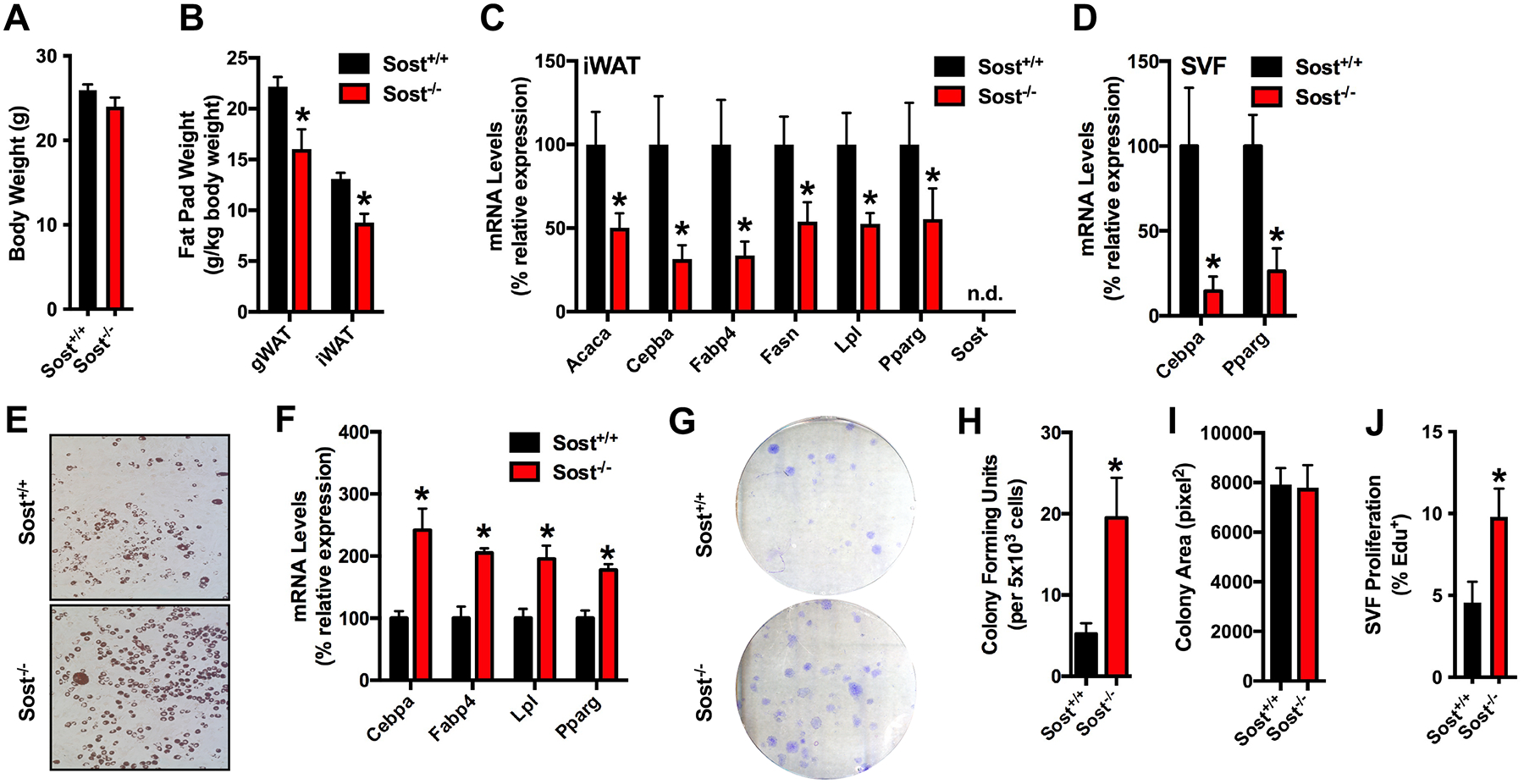
Figure 1. The stromal vascular fraction isolated from Sost−/− mice exhibits a paradoxical increase in adipocyte differentiation in vitro.

(A) Body weight of 8 week old male Sost+/+ and Sost−/− mice (n=7 mice/genotype). (B) Gonadal (gWAT) and inguinal (iWAT) fat pad weights (n=7 mice/genotype). (C) qPCR analysis of mRNA samples isolated from the inguinal fat pad of 8 week old mice (n=6 mice/genotype). (D) qPCR analysis of mRNA samples from stromal vascular fraction (SVF) cells freshly isolated from the inguinal fat pad (n=4–6 mice/genotype). (E and F) In vitro differentiation of SVF cells isolated from the inguinal fat pad of 8 week old male Sost+/+ and Sost−/− mice was assessed by Oil Red O staining (E) and qPCR analysis (F, n=4–6 mice/genotype). (G-I) Colony forming capacity and colony size were assessed by seeding 5×10³ SVF cells per 100mm plate (n=10–11 mice/genotype). (J) EdU incorporation was assess as a marker of proliferation (n=6–7mice/genotype). All data are represented as mean ± SEM. *p < 0.05.
