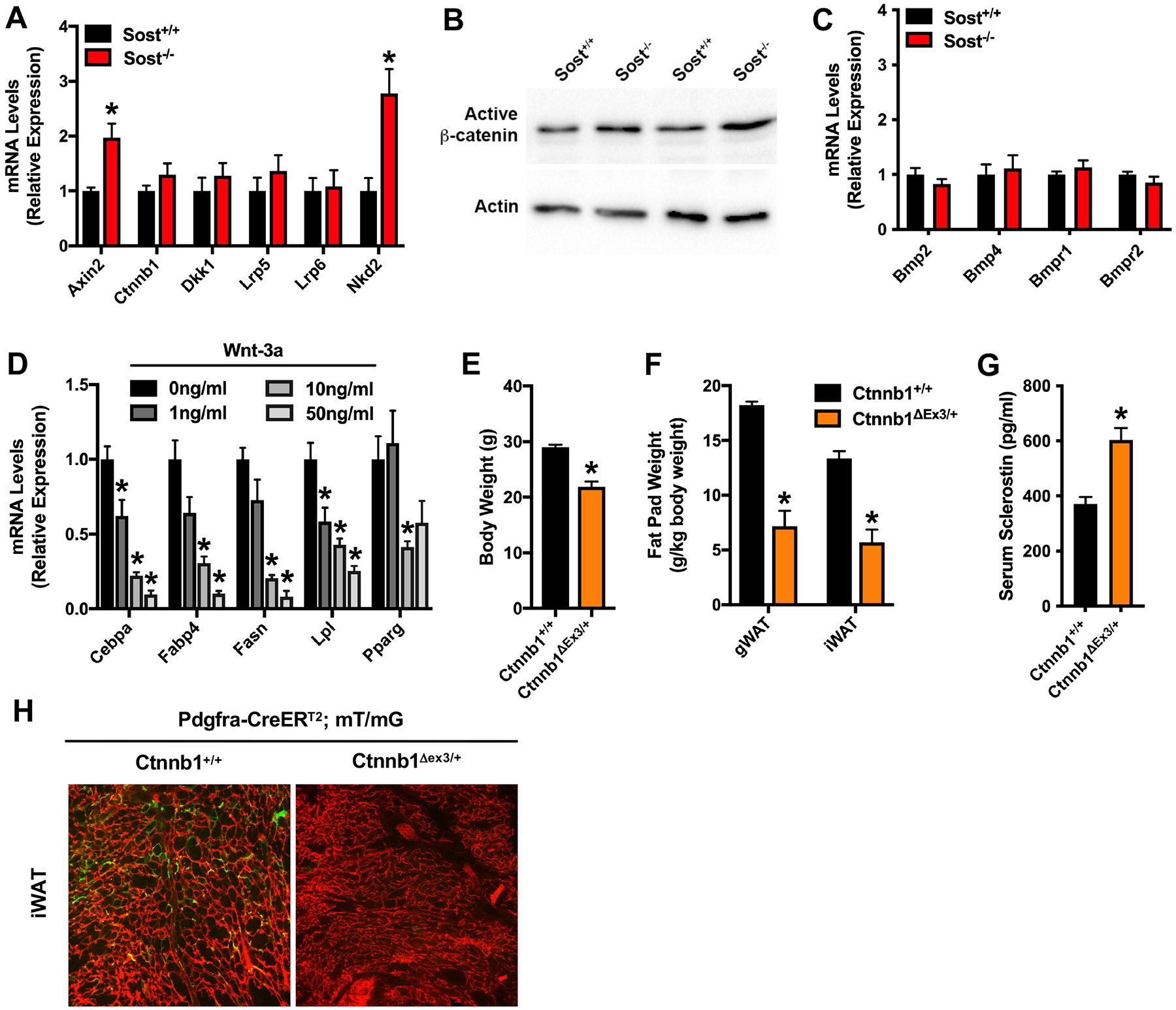
Figure 3. Wnt signaling inhibits APC differentiation.

(A) qPCR analysis of Wnt-related gene expression in CD45−: Sca1+: PDGFRα+ APCs (n=6mice/genotype). (B) Western blot analysis of active, non-phosphorylated β-catenin in APCs. Analysis for two lysates is shown for each genotype. (C) qPCR analysis of BMP-related gene expression in CD45−: Sca1+: PDGFRα+ APCs (n=6mice/genotype). (D) Adipogenic gene expression in CD45−: Sca1+: PDGFRα+ APCs treated with 0–50ng/ml recombinant mouse Wnt-3a (n=6/treatment group). (E and F) Body weight (E) and fat pad weight (F) in Pdgfra-CreERT2; mT/mG mice that contain wild type Ctnnb1 alleles (Ctnnb1+/+) or a Cre-inducible, constitutively active mutant allele (Ctnnb1Δex3/+) (n=5–7mice/genotype). (G) Serum sclerostin levels measured by ELISA in Ctnnb1+/+ and Ctnnb1Δex3/+ mice (n=5–7mice/genotype). (H) Representative fluorescent micrographs of mRFP and mGFP expression in the iWAT from lineage tracing studies with Ctnnb1Δex3/+; Pdgfra-CreERT2; mT/mG and control littermates (n=5–7mice/genotype). All data are represented as mean ± SEM. *p < 0.05.
