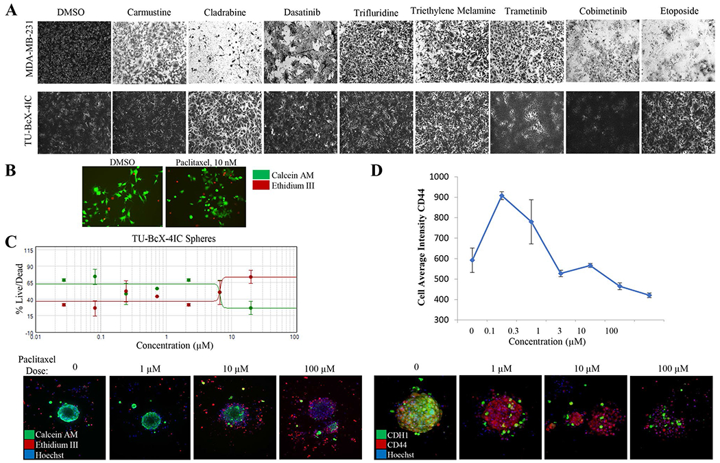
Fig. 4.

Chemosensitivity profiles of TU-BcX-4IC cells. A TU-BcX-4IC-derived cells and MDA-MB-231 cells were treated with the NCI-approved oncology drug panel for three days. Overall, TU-BcX-4IC cells were more chemoresistant than MDA-MB-231 cells. B TU-BcX-4IC adherent cells were resistant to paclitaxel treatment (10 nM, 72 h), observed with a live/dead Calcein-AM/EthD III fluorescent stain. Green = live cells, Red = dead cells. C U-BcX-4IC organoids were treated with varying doses of paclitaxel (1 μM, 10 μM, 100 μM) and stained with the live/dead cytotoxicity stain and Hoechst nuclear stain using the Pu·MA system (Protein Fluidics, Burlingame CA) and images were obtained using confocal microscopy (Molecular Devices, San Jose CA)
