
Fig. 6.
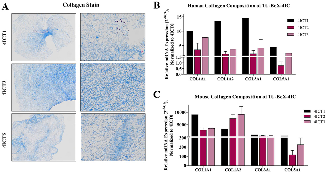
Collagen composition of serially transplanted TU-BcX-4IC PDX tumors. Serially transplanted TU-BcX-4IC PDX tumors were decellularized and consecutive passages of TU-BcX-4IC tumor serially transplanted in mice (T1, T3, T5) are exhibited. A Masson’s Trichrome was used to stain decellularized tumors to visualize collagen fiber organization. Decellularized tumors were formalin fixed, paraffin embedded, stained and representative images are shown at 10 × and 100 × magnification. COL1A1, COL1A2, COL3A1 and COL5A1 were analyzed using qRT-PCR with both B human- and C mouse-specific primers. Represented data is normalized to mouse-specific primers to evaluate relative gene expression. Due to the limited availability of tumor tissue, analyses were performed in duplicate, except for T1 and T6 which were single samples only. Error bars represent S.E.M. *p < 0.05, ***p < 0.001