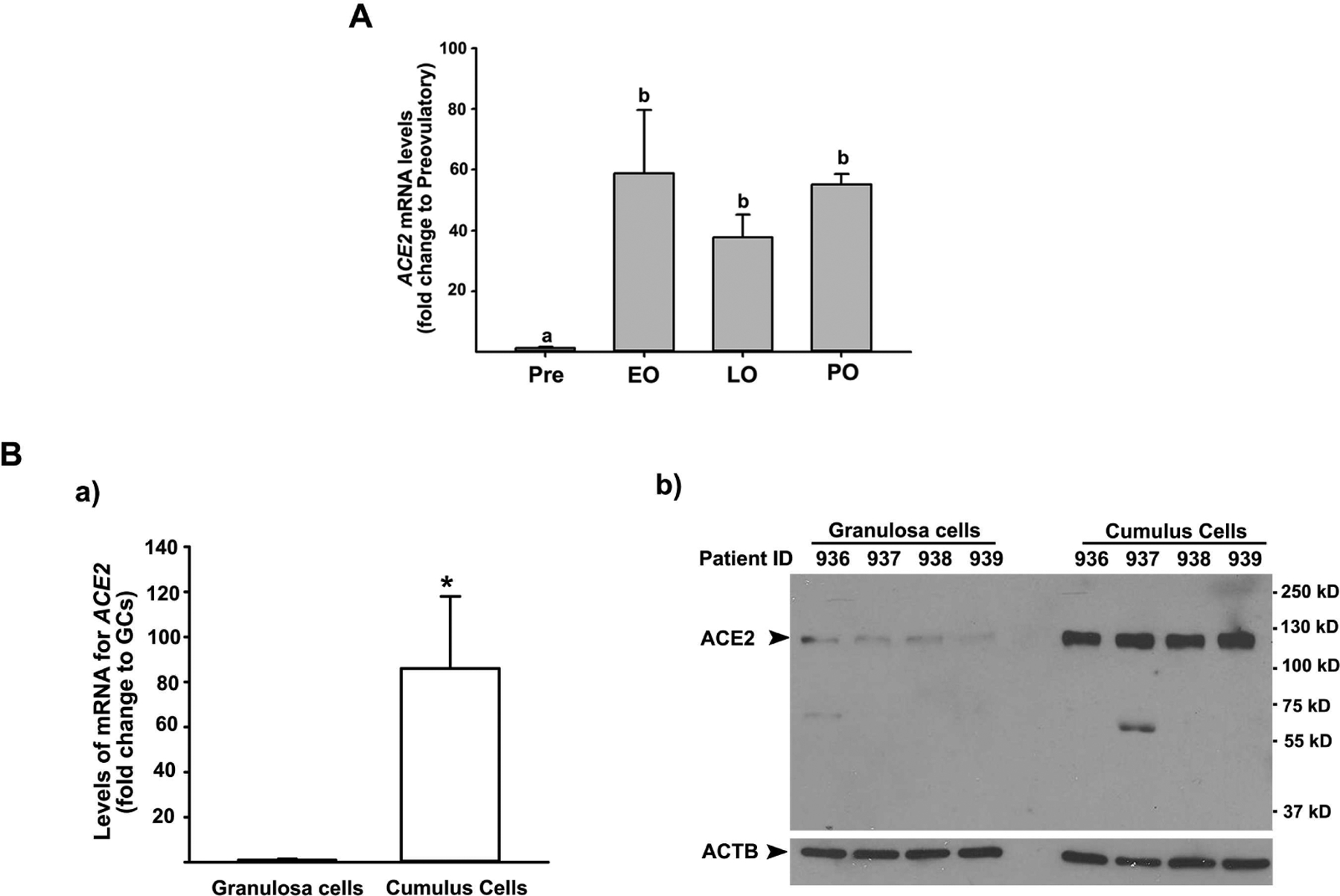
Figure 1. The levels of mRNA for ACE2 in granulosa cells and cumulus cells of periovulatory follicles.

A) Dominant follicles were retrieved from the ovaries of women undergoing laparoscopic tubal sterilization before the LH surge or at various times after recombinant hCG administration and divided into four phases: pre- (Pre, n=6), early (EO, n=5), late (LO, n=6), and post- (PO, n=2) ovulatory phases. The levels of mRNA for ACE2 were measured using qPCR in granulosa cells isolated from a dominant follicle collected at Pre, EO, and LO and whole follicles retrieved at PO and normalized to the levels of GAPDH mRNA in each sample. The levels were presented as fold change to Pre values. Bars with no common superscripts are significantly different (p < 0.05). B) Cumulus cells and granulosa cells were collected at the time of oocyte retrieval from women undergoing a standardized IVF procedure. a) The levels of mRNA for ACE2 were measured by qPCR and normalized to the levels of RNA18S5 in each sample (n = 5 independent samples). * p < 0.05. b) A representative Western blot image detecting ACE2 protein. Samples loaded were from independent patients indicated. ACTB detection in each lane was used as a protein loading control.
