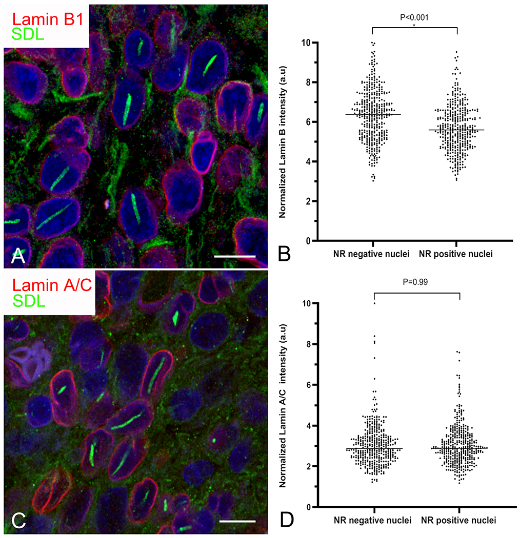
Figure 5:

Rods are associated with selective depletion of lamin B1. A, C) Confocal images of an oligodendroglioma showing visibly reduced lamin B1 (A; red) but not lamin A/C (C; red) staining intensity in cells with rods (green). Bars=10 μm. B, D) Grouped scatter plots of pooled data from astrocytomas and oligodendrogliomas showing significantly reduced staining intensity for lamin B1 (B) but not lamin A/C in cells with nuclear rods (NR).
