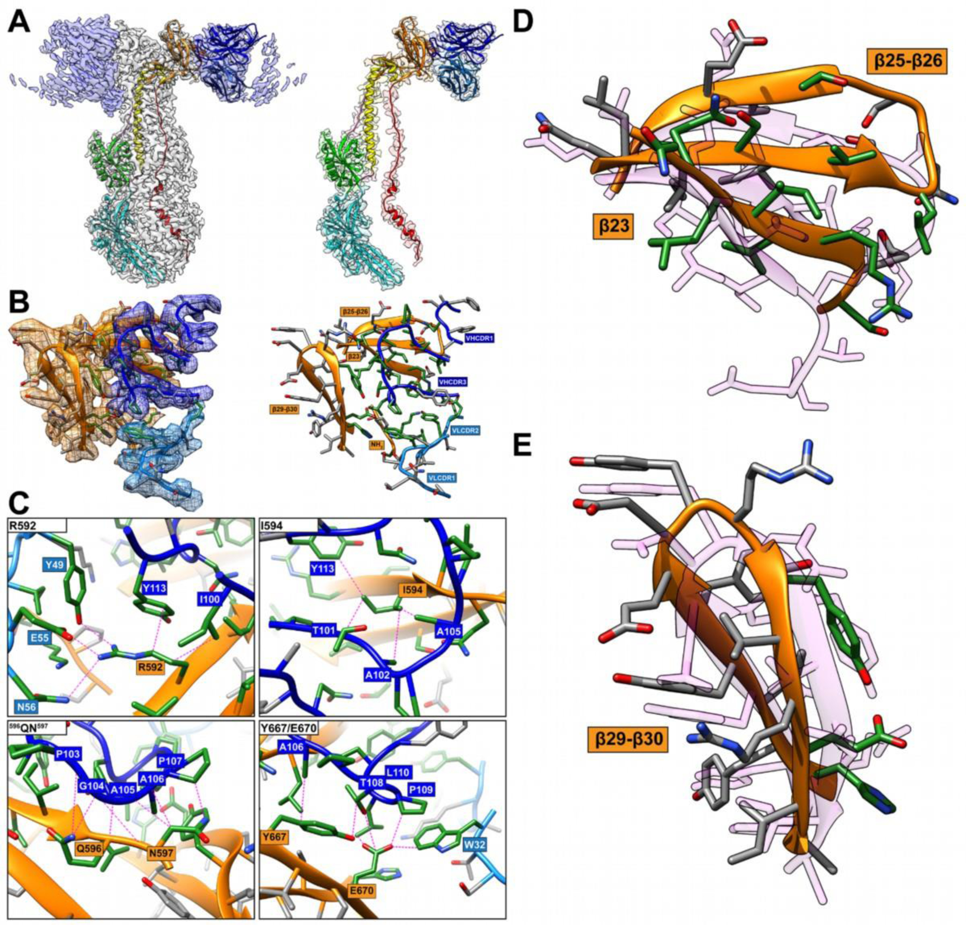
FIGURE 1.

Comparison of the RaptorX model to critical amino acid side chains identified at the gB-93k interface. Images A to C were reproduced from Oliver et al., 2020 under the Creative Commons Attribution 4.0 International license. A, Near-atomic resolution (2.8Å) cryo-EM structure of human neutralizing mAb 93k Fab fragments bound to native VZV gB. The left panel shows the cryo-EM map gB trimer (grey) and the 93k Fab fragments (blue) with the underlying model for one gB protomer and a 93k Fab fragment. Domains are colored accordingly: DI (cyan), DII (green), DIII (yellow), DIV (orange), DV (red), linker regions (hot pink), 93k VH (blue) and 93k VL (light blue). The left panel shows a segmentation of the cryo-EM map for one VH and VL chain of a 93k Fab fragment bound to a protomer of VZV gB. The structures of VZV gB and 93k VH and VL are represented as ribbons. B, Extracted density (left panel) for the gB-93k interface. The densities of gB DIV (orange), 93k VH chain (blue) and 93k VL chain (light blue) are highlighted. A ribbon diagram and side chains of the amino acids at the extracted densities are shown with those highlighted in green representing the interactions formed at the gB-93k interface. The right panel duplicates the left panel but without the extracted cryo-EM map densities. The β23, β25–26, β29–30 and the NH2 terminus of gB are highlighted with orange boxes, and the VHCDR1, VHCDR3, VLCDR1 and VLCDR2 are highlighted by blue boxes; VH – dark blue, VL – light blue. C, Molecular interactions between gB and the VH and VL chains of mAb 93k. The four panels show the interactions between gB residues R592, I594, 596QN597 and Y667/E670D with mAb 93k. Dotted lines (magenta) represent molecular interactions calculated using Find Contacts (Chimera). D and E, Comparison of RaptorX model T1036s1TS487_1-D1 to the cryo-EM structure of β23, β25–26 and β29–30 where mAb 93k binds to VZV gB. The RaptorX model is shown in pink.
