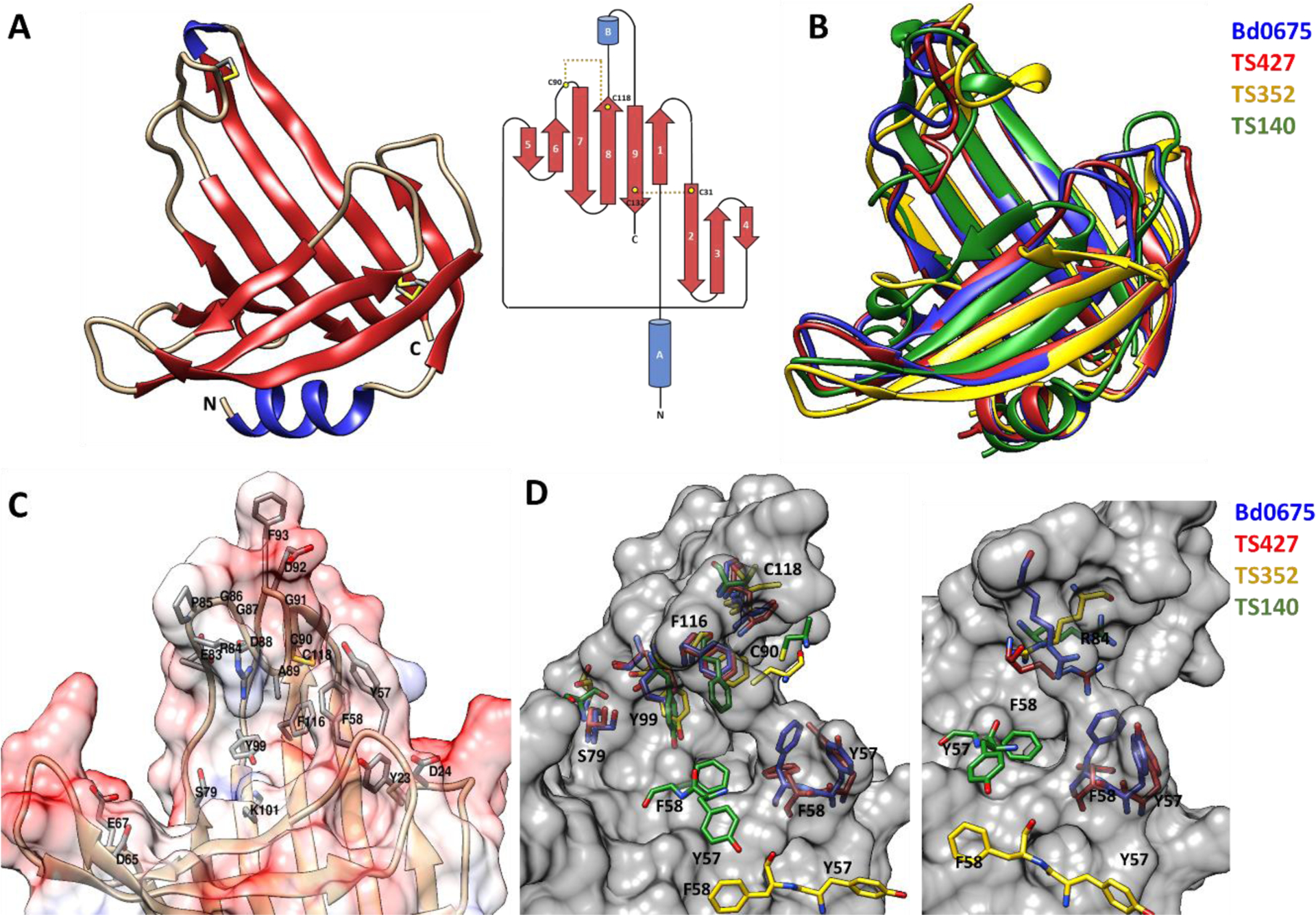
FIGURE 10.

A, Crystal structure and topological diagram of Bd0675 showing a β-roll like architecture stabilised by two disulphide bonds (in yellow). B, Superposition of Bd0675 crystal structure (blue) and the three best models: TS427 (red), TS352 (yellow) and TS140 (green). C, Surface potential representation of the putative ligand binding cleft. The proposed ligand binding groove of Bd0675 is formed mainly by acidic and hydrophobic residues. D, Comparison of the proposed ligand binding site of the Bd0675 crystal structure and the three best models.
