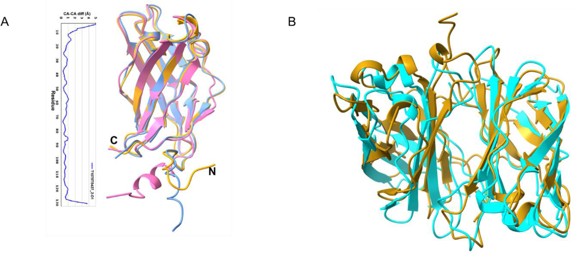
FIGURE 11.

A, Superposition of the Tsp1 monomer crystal structure (golden yellow) with top two CASP14 models T1078TS427_2-D1 (blue) and T1078TS314_1-D1 (pink). Also, given here is the plot of CA-CA distance (in Å) between crystal structure and CASP14 model T1078TS427_2-D1. B, Comparison of dimeric structure of Tsp1 (golden yellow) with the best predicted multimeric model (T1078TS177_3o, cyan).
