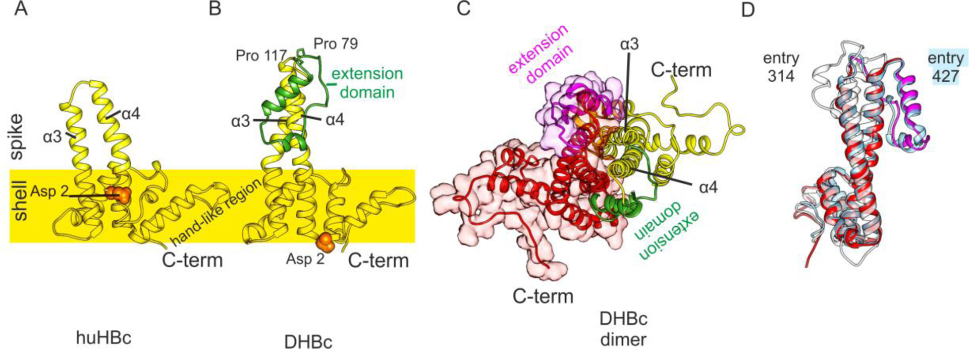
FIGURE 13.

Structures of DHBc PDB: 6YGH120 and huHBc PDB: 6HTX115. A, View of the huHBc monomer perpendicular to the dimer axis. The approximate position of the capsid shell is indicated to mark the protruding part of the spikes. Each monomer contributes the helices α3 and α4 to the spike and dimer interface. The C-terminus and the hand-like region mark the inter-dimer contact sites. The N-terminus (Asp2, orange spheres) is part of the intra dimer interface in huHBc (A) but not in DHBc (B). B, View of the DHBc monomer perpendicular to the dimer axis. The extension domain is delineated by Pro79 and Pro119 and is shown in green. C, View of the DHBc dimer perpendicular to the dimer axis. The other monomer is shown in red/magenta. The two extension domains (magenta and green) are part of the dimer interface and broaden the core spike. D, View onto the dimer interface of one monomer (red/magenta in C), with the superimposition of the two best scoring predicted models (entry 427, group AlphaFold2 in light blue; entry 314, group FEIG-R1 in white.
