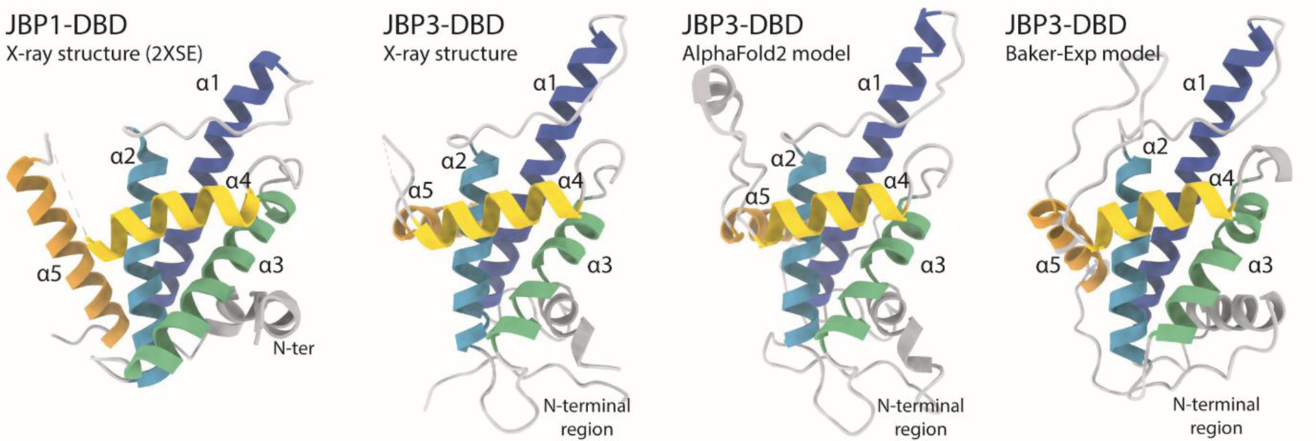
FIGURE 9.

Structures of J-DNA binding domains. From left to right: the structure of JBP1-DBD is the closest homologue to the CASP14 target crystallographic structure of JBP3-DBD that is shown next to it; the AlphaFold2 model has all independent evidence in essentially identical orientation as the experimental structure; the second best model (from the Baker group) shows a different orientation for the α5 helix and the N-terminal region.
