
Fig. 1.
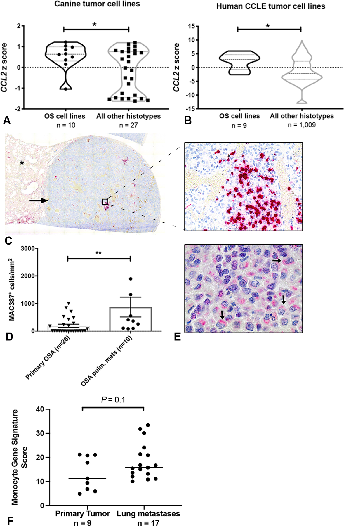
Canine and human osteosarcoma cells express CCL2 and are enriched for monocytes within pulmonary metastases. (A) CCL2 mRNA expression in canine OS cells, as compared to all cell lines of non-osteosarcoma histo-type. *P = 0.04, unpaired two-tailed Student’s t test. (B) CCL2 mRNA expression in human OS cells as compared to all cell lines of non-osteosarcoma histo-type within the Broad Institute Cancer Cell Line Encyclopedia (CCLE). *P = 0.01, unpaired two-tailed Student’s t test. (C) Sub-gross overview and corresponding 400x magnification image of a canine osteosarcoma pulmonary metastasis containing extensive intra-tumoral infiltrates of MAC387+ monocytes and macrophages (cells labeled in red; asterisk denotes normal lung parenchyma). (D) 1,000x magnification image of the same metastatic lesion shown in (D) demonstrating strong cytoplasmic, peri-nuclear positive immunolabeling of canine OS tumor cells for CCL2 (arrows). (E) Quantitative image analysis of MAC387+ myeloid cells in osteosarcoma pulmonary metastases as compared to primary tumors (n=10 and 26 animals per group, respectively). **P = 0.004, two-tailed Mann Whitney test, data plotted as mean ± SEM. (F) Mean expression of monocyte immune signature genes in human osteosarcoma pulmonary metastases vs. primary tumors using RNAseq data obtained from CC Wu et al. P = 0.1, unpaired two-tailed Student’s t test.