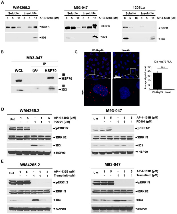
Figure 4: ID3 is a novel client protein of HSP70.
(A) Western blot probed with indicated antibodies of detergent-soluble and -insoluble lysates from WM4265.2, M93-047 and 1205Lu cell lines treated with 0, 5 and 10 μM AP-4-139B for 24 hours. (B) Lysates from M93-047 cells were immunoprecipitated with IgG or anti-HSP70 antibodies and probed for ID3. WCL= whole cell lysate. (C) Proximity ligation assay for HSP70-ID3 complexes in M93-047 cells. Individual HSP70-ID3 interactions are visualized by fluorescent signal (red) with nuclei counterstained with DAPI. Scale bar = 50 μm. Representative images are maximum intensity projects from z-stacks. Right panel: quantification of the HSP70-ID3 interactions measured as the average number of PLA signals per nuclei, from > 100 cells analyzed from random fields in each of two technical replicates. *** p-value < 0.001, assessed by two tailed student’s t-test. (D and E) Western blot probed with indicated antibodies of lysates from WM4265.2 and M93-047 cell lines treated with the indicated concentrations of AP-4-139B, PD901 and/or Trametinib for 24 hours.

