Fig. 5.

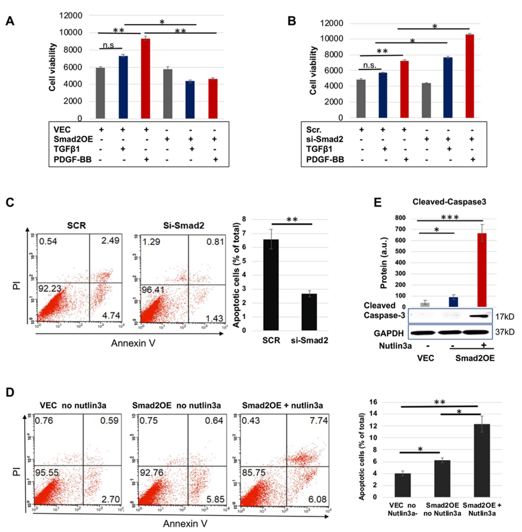
p53 activation potentiates Smad2’s pro-apoptotic function.
A and B. AoSMC viability assay (CellTiter-Glo).
C and D. Apoptosis assay (FACS). Black bars represent averaged values based on three independent repeat experiments (mean ± SD, n = 3). The apoptotic cells include Q2 and Q4, late- and early-phase apoptotic populations, respectively. The Flow Jo software was used.
E. Apoptosis assay (cleaved caspase3).
Prior to assays, human primary aortic SMCs (AoSMCs) were cultured, transfected, and cytokine-treated, as described for Fig. 1. Quantification was performed as described for Fig. 1; mean ± SD, n = 3 repeats.
Statistics: One-way ANOVA/Bonferroni post-hoc test; Student’s t-test was performed in C; *P < 0.05, **P < 0.01, ***P < 0.001; n.s. not significant.
