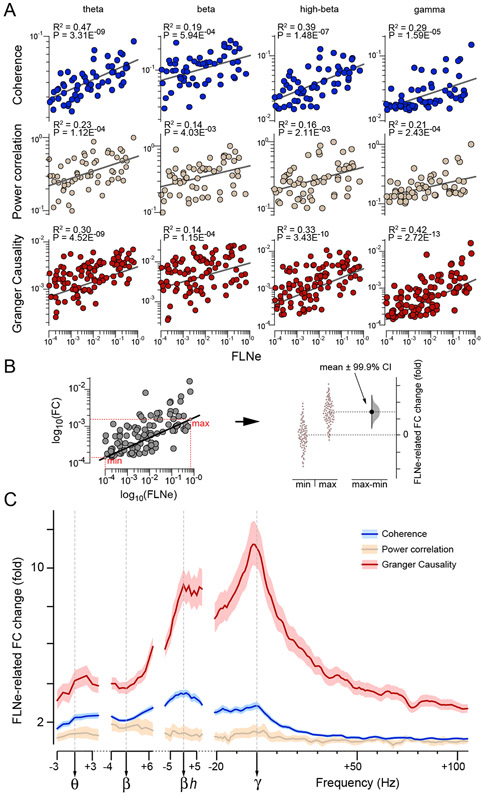
Figure 5. FC and AC display frequency-dependent covariance.
(A) Scatterplots between the three FC types (indicated to the left of the rows) and FLNe. For coherence and power correlation, each dot corresponds to a pair of areas, for which the combined FLNe in both directions was based on more than 10 labeled neurons (N=60). For GC, each dot corresponds to an anatomical projection, for which the FLNe in the same direction as the corresponding GC was based on more than 10 labeled neurons (100). FC values were averaged over monkeys before correlation analysis. Note logarithmic scaling on x- and y-axes.
(B) With both axes in log10 units, subtraction of FC values between minimum and maximum AC values (left), can be interpreted as FLNe-related fold-change of FC (right).
(C) FLNe-related FC change as a function of FC frequency. Log10(FC) spectra (color coded, legend top-right) have been aligned to individual peak frequencies before averaging over monkeys and then correlated with log10(FLNe). Mean over all trials ±99.9% confidence intervals from bootstrap estimates over trials.
See also Figure S5.

