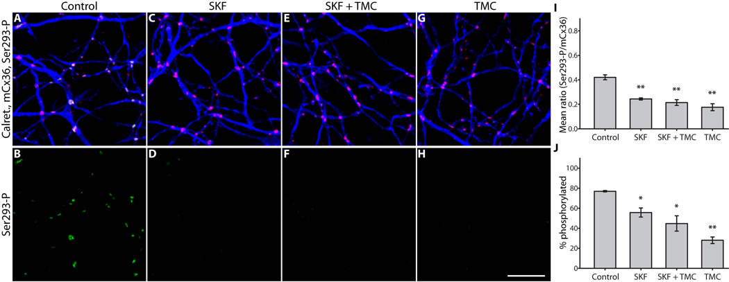
Figure 5. PP1 negatively regulates the dephosphorylation of Cx36.
A & B, Control; color scheme and antibodies are the same as Figure 2. C & D, D1R activation (SKF38393, 10 µM) greatly diminished Ser293-P labeling. E & F, Inhibition of PP1 (tautomycetin, 10 nM) did not alter the effects of D1R activation. G & H, Inhibition of PP1 alone was sufficient to cause a strong reduction in Ser293-P labeling. I, Summary of data shows that inhibition of PP1 did not prevent the reduction in Ser293 phosphorylation caused by D1R activation, and that PP1 inhibition alone significantly reduced Ser293 phosphorylation. J, Summary of data shows that changes in the percentage of Cx36 plaques that show detectable Ser293-P labeling follows the same pattern established for relative Ser293-P measurements in I. Error bars are s.e.m, n = 4. Asterisk denotes P < 0.05, two asterisks denote P < 0.01. Images are 1 µm-deep stacks. Scale bar in H is 10 µm.

