Figure-1:

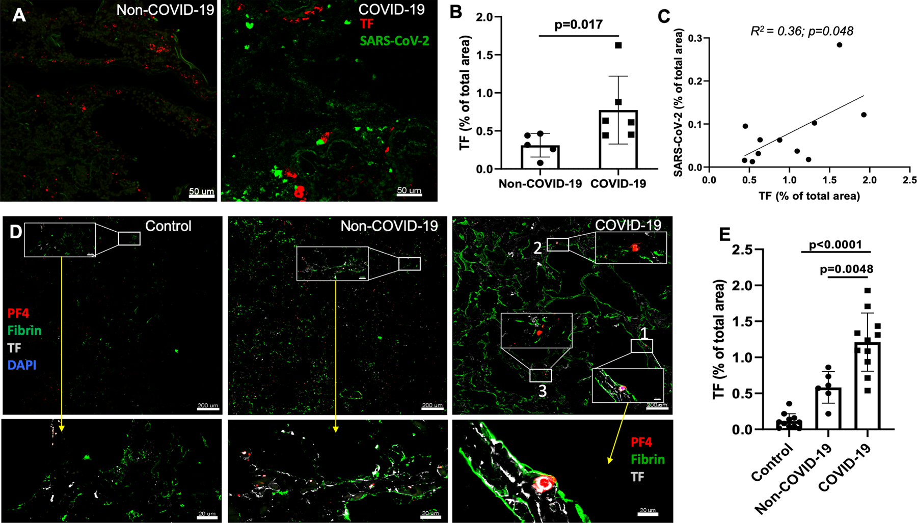
In situ hybridization detecting SARS-CoV-2 viral RNA and TF RNA. (A) Dual RNA in situ hybridization of SARS-CoV-2 (green color dots) and TF (red color dots) in COVID-19-ARDS and non-COVID-19 ARDS cases. (B) Quantification of TF RNA expression from RNAscope (n=6) using ImageJ program. (C) Correlation of TF expression with SARS-CoV-2 expression (combined RNAscope (n=6) and immunofluorescence (n=5); R2=0.66; p<0.01). Staining of TF mRNA and immunofluorescence produced similar results, and so the combined TF mRNA expression from 5 COVID-19 patients and TF staining from 6 COVID patients were shown. (D) Immunofluorescence assessment of lungs tissues from COVID-19-ARDS cases identifies higher TF expression associated with fibrin- and PF4-positive thrombi. Immunostaining for TF (white), fibrin (green) and PF4 (red) of COVID-19-ARDS and non-COVID-19 ARDS as well as pathologically normal control lung tissues. Inset with white box and bottom panels showing magnified vessels/area with fibrin- and/or platelet-rich thrombi. (E) Quantification of TF expression.
