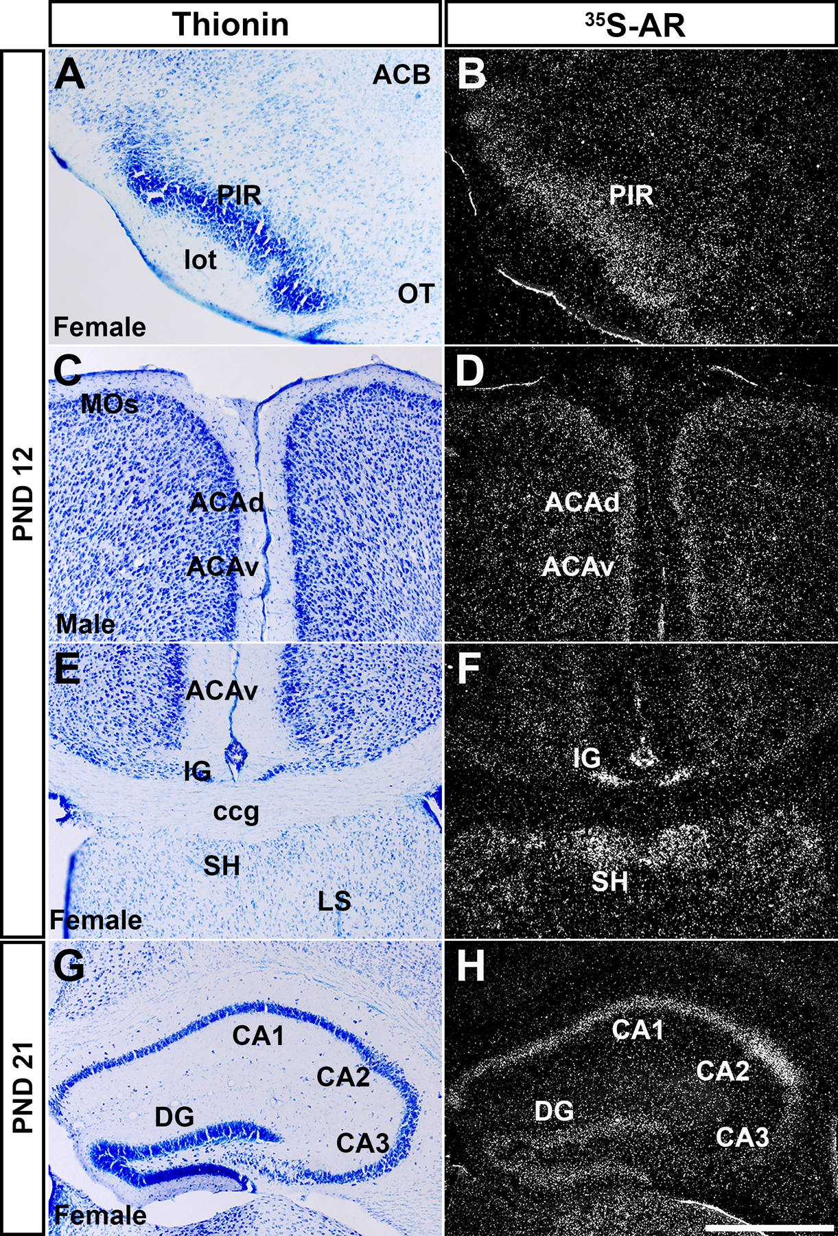
Figure 4: Ar mRNA expression in cerebral cortex in prepubertal male and female mice.

Images showing thionin staining for neuroanatomical reference (left column), silver grains corresponding to Ar mRNA (right column). Low Ar expression was observed in the piriform area (PIR, A-B), dorsal and ventral anterior cingulate area (ACAd and ACAv, C-D), induseum griseum, septohippocampal nucleus (IG and SH, E-F), and CA3, and high in field CA1 and CA2 (G-H). Abbreviations: ACB, nucleus accumbens, ccg, genu of corpus callosum, DG, dentate gyrus, lot, lateral olfactory tract, LS, lateral septal nucleus, MOs, secondary motor area, OT, olfactory tubercle. Scale bar = 200 µm.
