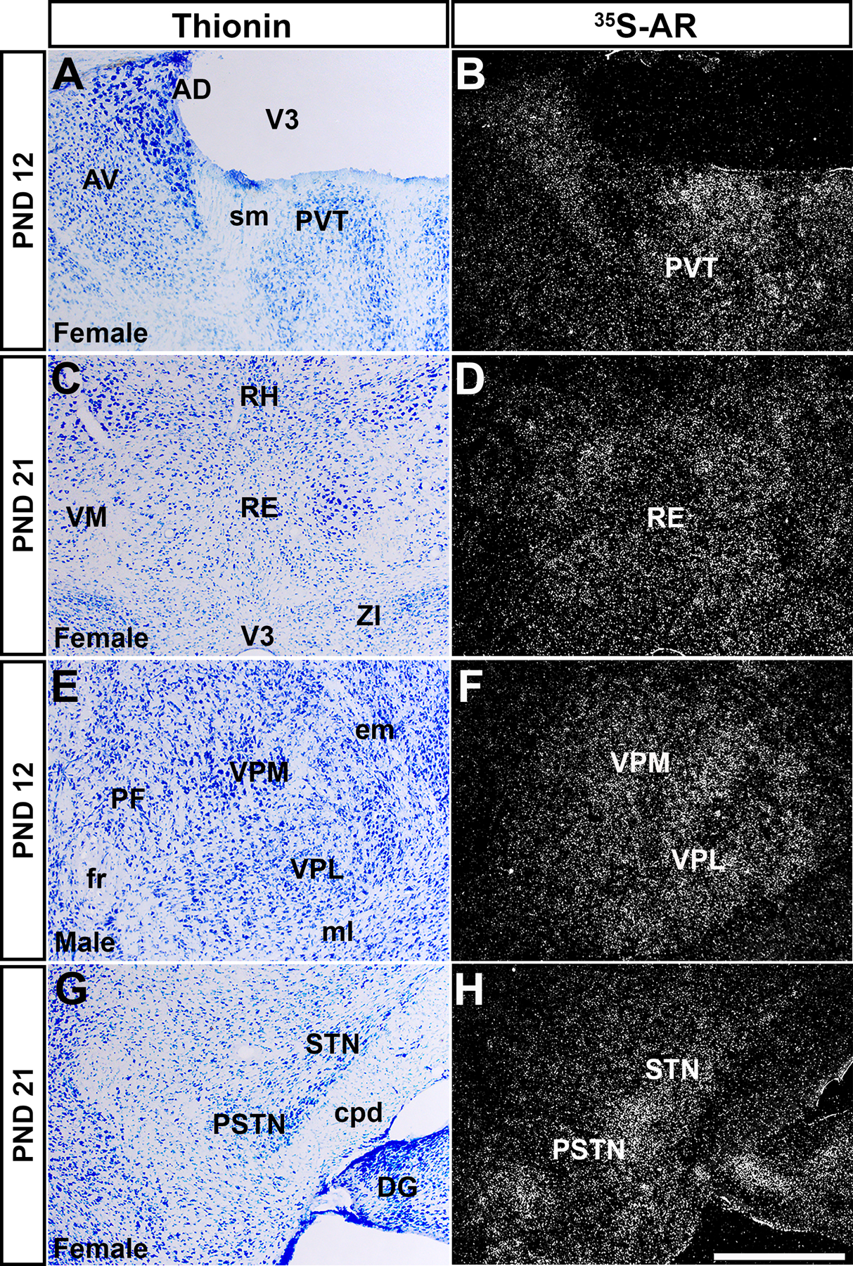
Figure 6: Ar mRNA expression in thalamic nuclei of male and female prepubertal mice.

Images showing thionin staining for neuroanatomical reference (left column), silver grains corresponding to Ar mRNA (right column). (A-B) Low silver grain deposition in the paraventricular nucleus of the thalamus (PVT), (C-D) low to moderate in the nucleus of reuniens (RE), (E-F) ventral posterolateral and posteromedial nuclei of the thalamus (VPL and VPM), (G-H) subthalamic and parasubthalamic nuclei (STN and PSTN). Abbreviations: AD, anterodorsal nucleus of the thalamus, AV, anteroventral nucleus of the thalamus, cpd, cerebral peduncle, DG, dentate gyrus, em, external medullary lamina of the thalamus, fr, fasciculus retroflexus, ml, medial lemniscus, PF, parafascicular nucleus, RH, rhomboid nucleus, sm, stria medullaris, VM, ventral medial nucleus of the thalamus, ZI, zona incerta. Scale bar = 200 µm.
