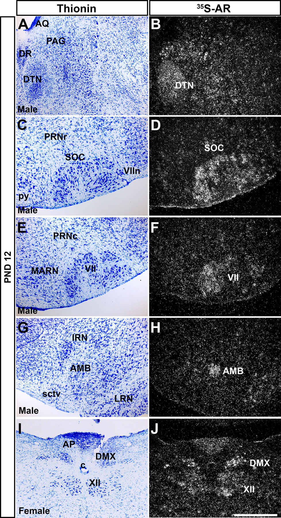
Figure 8: Ar mRNA expression in brainstem nuclei of prepubertal male and female mice.

Images showing thionin staining for neuroanatomical reference (left column), silver grains corresponding to Ar mRNA (right column). (A-B) Very low to low silver grain deposition in the periaqueductal gray (PAG), and low in the dorsal tegmental nucleus (DTN). (C-D) Low expression in the superior olivary complex (SOC), (E-F) facial motor nucleus (VII). (G-H) Moderate expression in the nucleus ambiguus (AMB). (I-J) Low to moderate expression in the dorsal motor nucleus of the vagus nerve (DMX) and hypoglossal nucleus (XII). Abbreviations: VIIn, facial nerve, AP, area postrema, AQ, cerebral aqueduct, c, central canal of the spinal cord/medulla, DR, dorsal nucleus raphe, IRN, intermediate reticular nucleus, LRN, lateral reticular nucleus, MARN, magnocellular reticular nucleus, PRNc, pontine reticular nucleus, caudal part, PRNr, pontine reticular nucleus, py, pyramid, sctv, ventral spinocerebellar tract. Scale bar = 200 µm.
