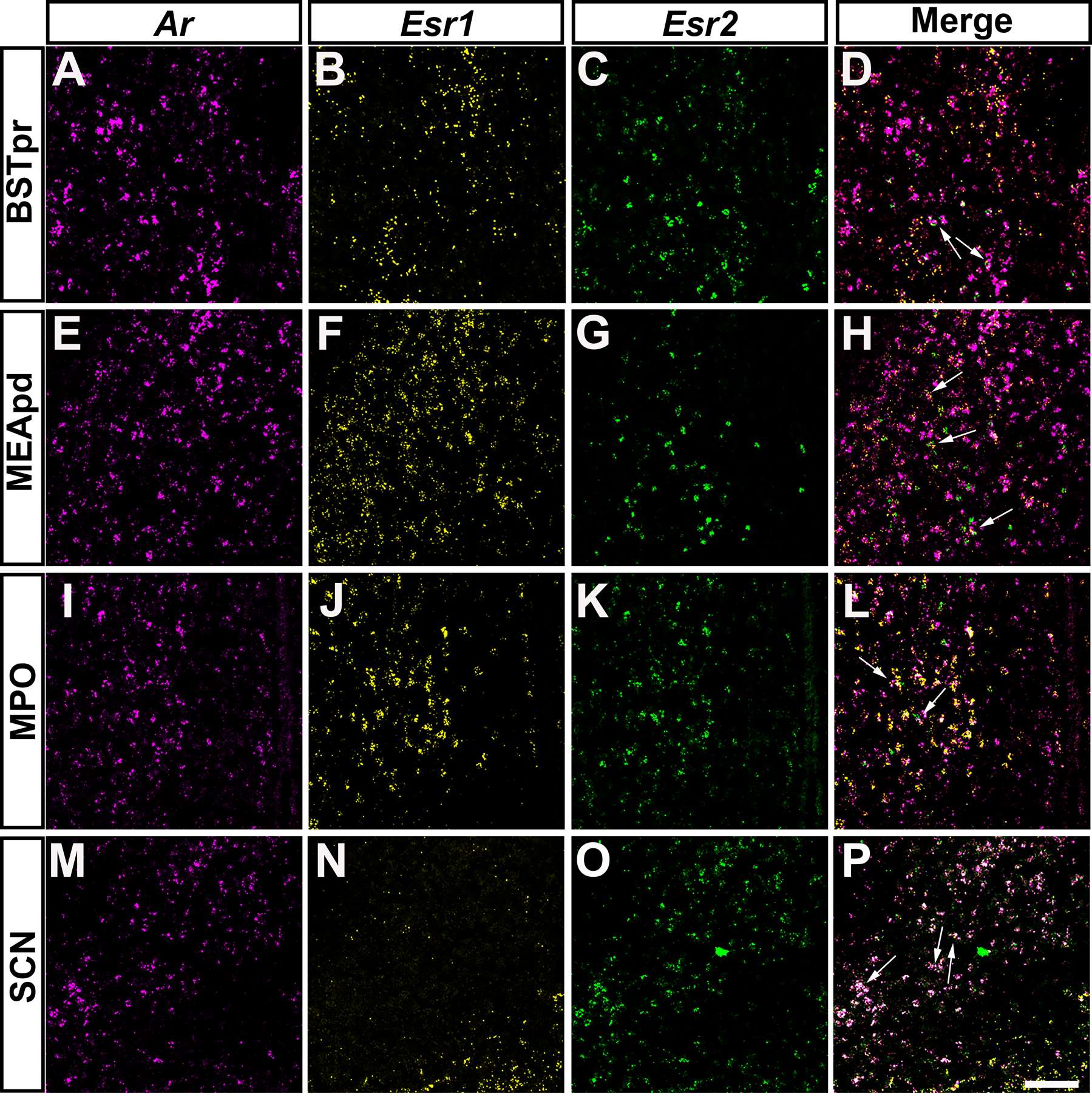
Figure 9: Ar mRNA expression overlaps with Esr1 and Esr2 in specific forebrain nuclei of prepubertal mice.

A-P, images showing fluorescent in situ hybridization signal for Ar (magenta, A, E, I, M), Esr1 (yellow, B, F, J, N), and Esr2 (green, C, G, K, O). Merge of all 3 channels shown in D, H, L, and P. Areas with Ar and Esr1 and/or Esr2 co-expression include the bed nucleus of the stria terminalis, principal nucleus (BSTpr, A-D), medial amygdalar nucleus, posterodorsal (MEApd, E-H), medial preoptic area (MPO, I-L), suprachiasmatic nucleus (SCH, M-P). Arrows show dual or triple-labeled neurons. Images shown are from postnatal day 12 (PND 12) female (BSTpr, MEApd, MPO) and male (SCH) mice. Scale bar = 100 µm.
