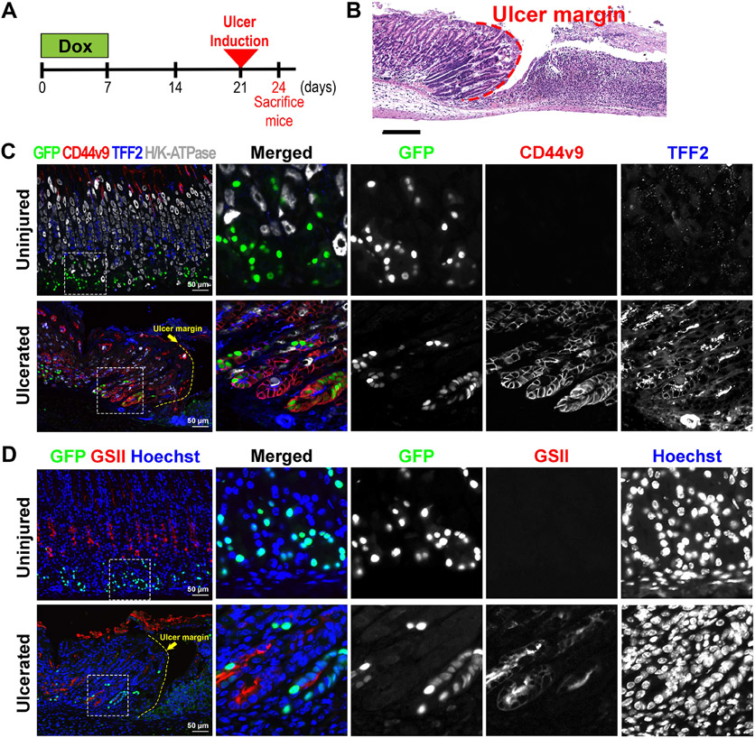
Figure 6. Lineage contribution of GFP-labeled chief cells in acetic acid-induced ulceration.
A) Scheme of acetic acid-induced ulceration in GIF-Cre-RnTnG mice. To first lineage label chief cells with GFP, mice received Dox for 1 week followed by 2 weeks off before the gastric ulceration was induced. The stomach tissues were collected 3 days after the ulceration. B) H&E-stained stomach sections from GIF-Cre-RnTnG mice at 3 days after acetic acid-induced injury. N = 3 mice. Dotted line indicates ulcer margin. C&D) Sections of the stomach tissues from uninjured or ulcerated lesions in the GIF-Cre-RnTnG mice were immunostained with (C) antibodies against GFP (green), CD44v9 (red), TFF2 (blue) and H/K-ATPase (white) and (D) antibodies against GFP (green) and GSII (red). Nuclei were counterstained with Hoechst. White dotted boxes depict enlarged regions and yellow dotted line indicates ulcer margin.

