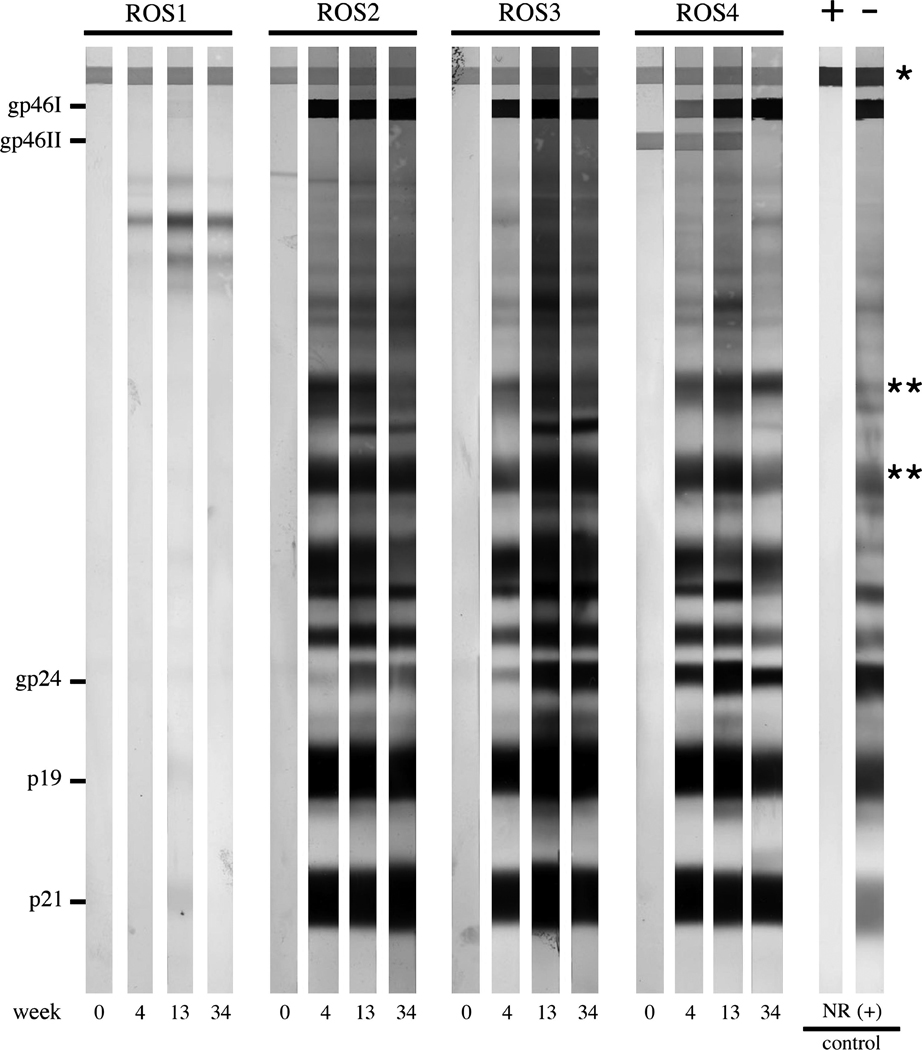
FIG. 3. Anti-HTLV-1 Western blot.

Representative results from selected animals are shown. ROS1 was inoculated with HTLV-1-negative Jurkat T lymphocytes, and never seroconverted. ROS-2, -3, and -4 were inoculated with HTLV-1-positive ACH.2 cells. All ACH.2-inoculated rabbits seroconverted by week 4 (gp46I, glycoprotein 46 HTLV-1 Env surface unit; gp46II, glycoprotein 46 HTLV-2 Env surface unit; gp24, HTLV-1 capsid; p19, HTLV-1 matrix; p21, HTLV-1 Env transmembrane unit; asterisks denote serum loading control bands, indicating comparable concentrations of serum immunoglobulin levels among the samples).
