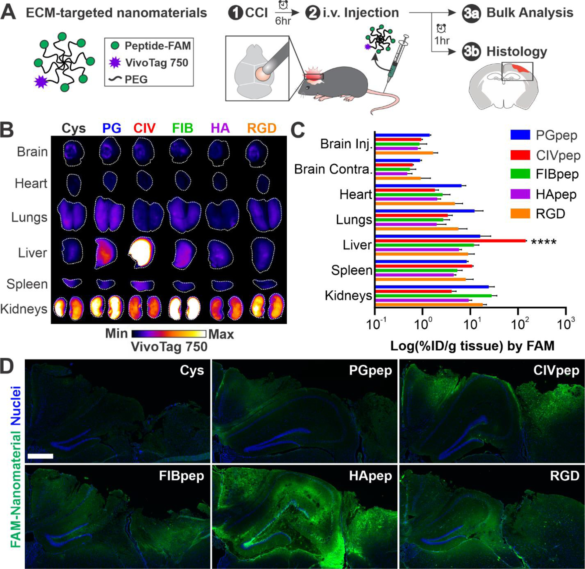
Figure 1.

Nanomaterial modification with HA-targeting peptide leads to widespread distribution in the injured brain after systemic administration. (A) Schematic of ECM-targeted nanomaterials and overview of experimental design. 6 hours post-CCI, ECM-targeted nanomaterials were intravenously administered. After 1 hour, organs were harvested for analysis of nanomaterial biodistribution and histology. (B) Surface imaging of VivoTag 750 from major organs of one representative mouse per nanomaterial (n = 3, white line indicates outline of organ). (C) Bulk quantification of percent injected dose nanomaterial per gram (% ID/g) tissue based on FAM fluorescence (n = 3, mean ± SEM, ****p ≤ 0.0001, two-way ANOVA and Tukey’s multiple comparisons post-hoc test within each organ group). (D) Representative images of the injured cortex in coronal brain slices (n = 3; blue, nuclei; green, FAM-labeled ECM-targeting peptide on nanomaterial; scale bar = 500 μm).
