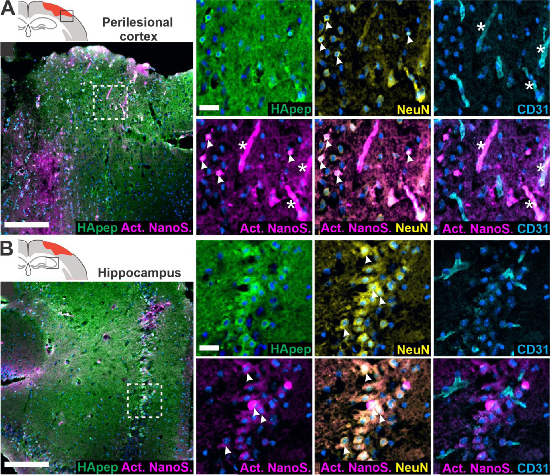
Figure 5.

Hyaluronic acid-targeted nanosensor activates within neuronal and endothelial cells in the perilesional cortex and hippocampus. Images from the (A) perilesional cortex and (B) hippocampus imaged for nanosensor distribution and activated nanosensor (box in schematic indicates imaging location). Immunostaining was performed for HApep (green, FAM), neurons (yellow, NeuN), and endothelial cells (cyan, CD31) (blue, nuclei; magenta, activated nanosensor; scale bar = 200 μm for larger images and 25 μm for insets). Colocalization of nanosensor activation with neurons or endothelial cells are denoted with arrows or stars, respectively.
