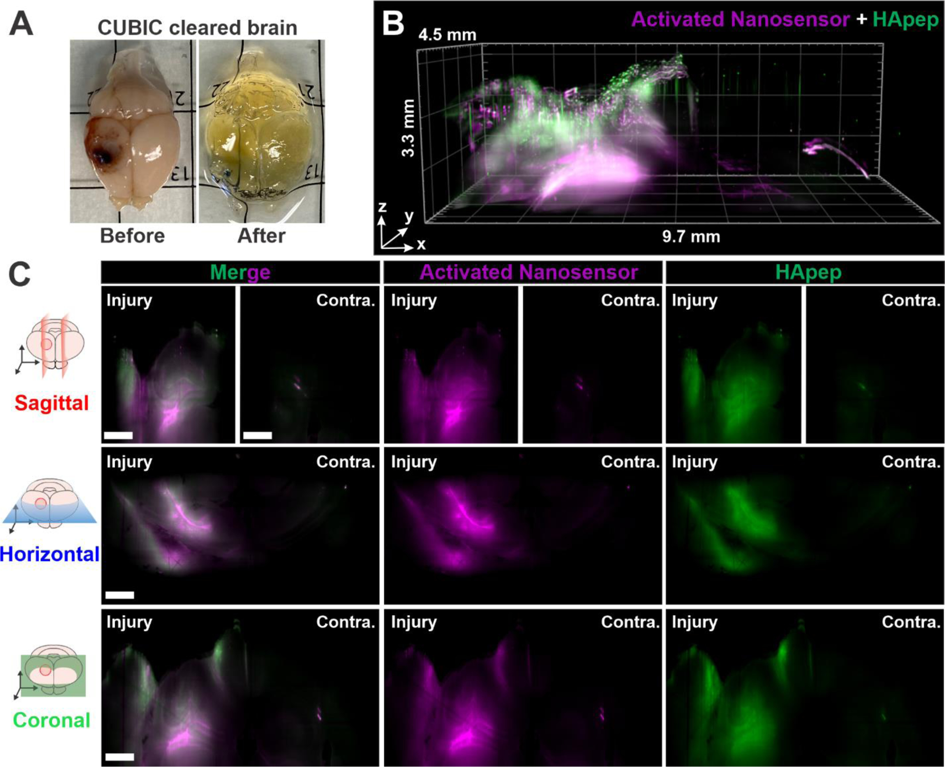
Figure 6.

Light sheet fluorescence microscopy (LSFM) of cleared tissues enables 3-dimensional (3D) reconstruction of nanosensor activation within the injured brain. (A) Image of injured brain before and after whole brain CUBIC clearing. (B) 3D render view. (C) Sagittal, horizontal, and coronal cross sections (magenta, activated nanosensor; green, HApep on nanosensor; scale bar = 1 mm; Contra. = contralateral hemisphere). See Supplementary Video and Supplementary Figure S10 for the clipping planes used to generate the cross-sections.
