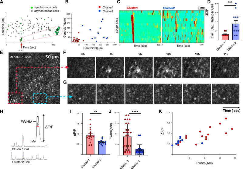
Figure 4. Characterization of spatiotemporally synchronous cells.
(A) Correlation between location and timing of individual cells and calcium elevation during synchrony event.
(B) Unsupervised k-mean clustering based on Euclidean distances reveals two categorical clusters of synchronous cells: those that are spatially localized (red) and those that are disperse (blue).
(C) Heatmap of single-cell fluorescence dynamics for clusters 1 and 2.
(D) Comparison of single-cell calcium elevation rate for clusters 1 and 2. Error bars represent standard deviations (unpaired t test, p < 0.001). (E) MIP spanning the time of high excess synchronicity (90–105 s).
(F and G) Localized region containing cluster 1 cells (F) compared with comparable-sized region (G).
(H–K) Comparison of normalized calcium-dependent fluorescence changes (ΔF/F) and FWHM of clusters 1 and 2. Error bars represent standard deviations (unpaired t test, p = 0.002 for I, p < 0.001 for J)

