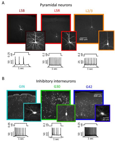
Figure 1. Six electrically and morphologically distinct types of neurons in mouse somatosensory cortex.
A. Biocytin-filled reconstructions (top) and intracellular recordings (bottom) showing the laminar location, morphology, and spiking responses typical of Layer 5 bursting (left, dark red), Layer 5 regular-spiking (center, red) and Layer 2/3 regular-spiking (right, orange) pyramidal neurons.
B. Biocytin-filled reconstructions (top) and intracellular recordings (bottom) showing the laminar location, morphology, and spiking responses typical of GFP+ inhibitory neurons in the GIN (left, cyan, adapting-spiking), G30/SZ (center, green, irregular-spiking), and G42 (right, blue, fast-spiking).

