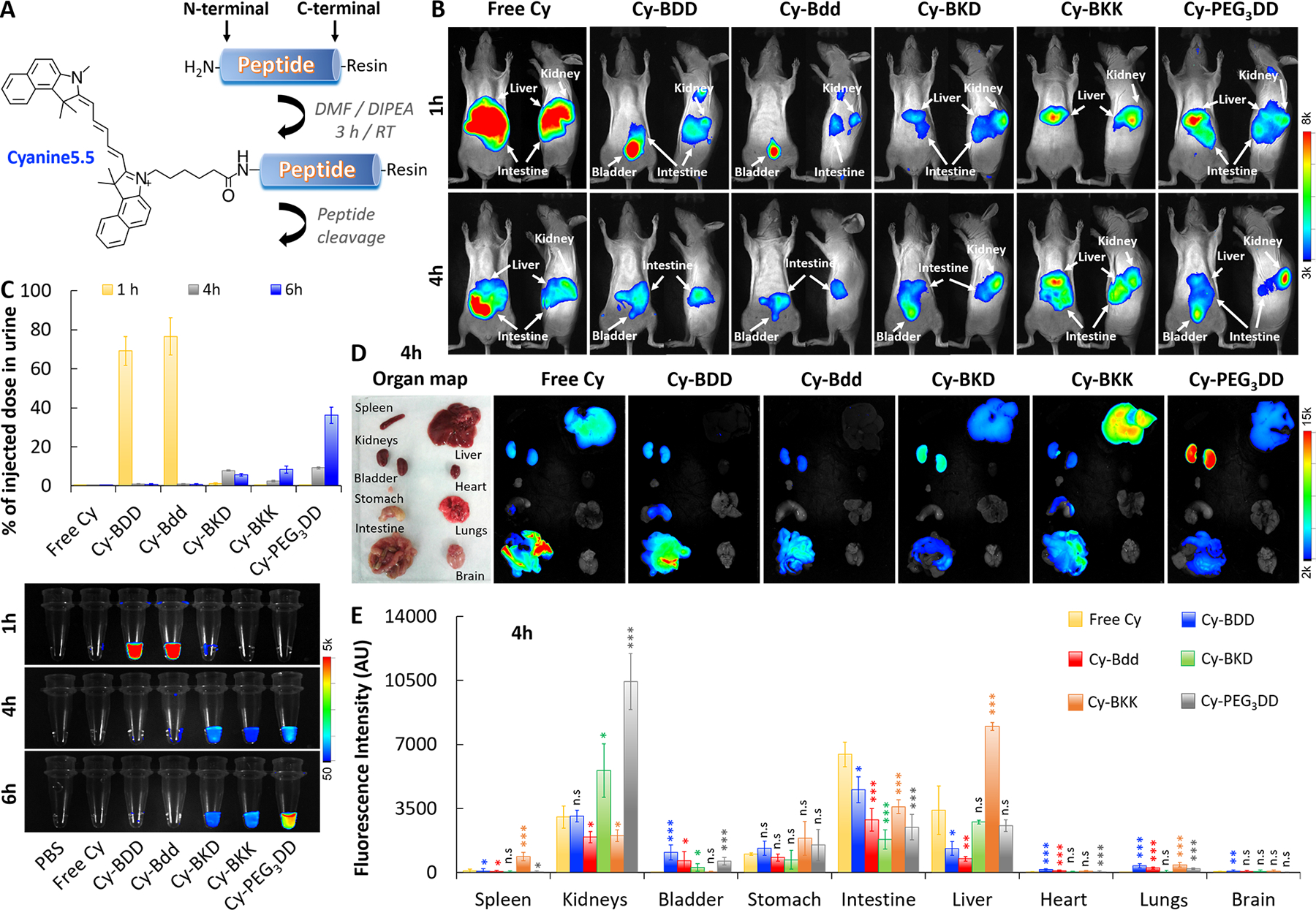
Fig. 2: Bdd effectively distributes the conjugated Cyanine5.5 fluorophore to the urinary system.

A, A synthetic scheme of the Cyanine5.5-labeled peptide (Cy-peptide) analogues. B, Representative merged fluorescence/white light-images of SHO mice acquired 1 and 4 h after tail-vein injection of the different Cy-peptide analogues (0.5 nmol, 150 μL) or free Cyanine5.5 (n=4/group). C, Plots comparing the amount of fluorophore in urine (% of injected dose) based on the measured fluorescence. (Lower panel) Fluorescence image of the urine samples (20 μL) collected from the same animals 1, 4, and 6 h after the Cy-peptides or fluorophore administration (n=4/group). For each time point, the bladders of the animals were completely emptied using a sterile 24 G pediatric venous catheter. D, Representative ex vivo merged fluorescence/bright light images of the organs harvested from animals 4 h after tail-vein injection of different Cy-peptides (n=4/group). E, Bar chart comparing peptide distribution in the harvested organs (n=4/group), based on the total fluorescence intensity. (Student’s t-test; *p<0.05, **p<0.01, and ***p<0.001).
