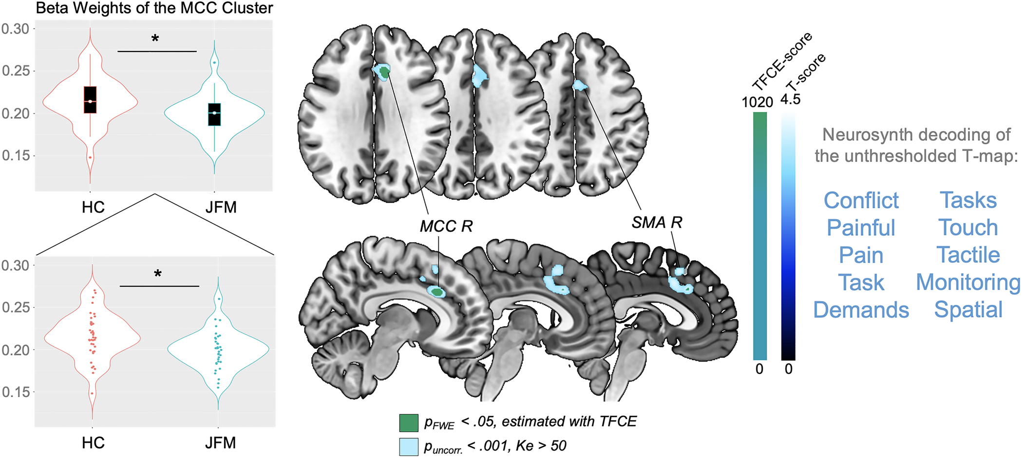
Figure 1. Regions of significantly reduced gray matter volume in adolescents with JFM.

Significant between-group gray matter volume differences (contrast: JFM < Controls). Results are presented at a significance level of pFWE-corr<.05, estimated with the TFCE approach (in green) and puncorr.<.001, Ke>50 voxels (in blue). At the right of the image, we display the 10 functional annotations most associated with the unthresholded t-map, as obtained with meta-analytic decoding using Neurosynth. FWE-corr: Family-Wise Error-corrected; HC: Healthy Controls; JFM: Juvenile Fibromyalgia; Ke: Cluster extent in voxels; L: Left; MCC: Anterior-Midcingulate Cortex; R: Right; SMA: Supplementary Motor Area; TFCE: Threshold-Free Cluster Enhancement; uncorr: uncorrected.
