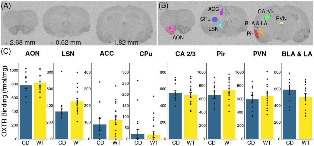
FIGURE 3.
No significant differences in oxytocin receptor density in Complete Deletion mice. (A) Coronal sections from a representative mouse brain used in iodinated ornithine vasotocin analog ([125I]OVT) autoradiography analysis with corresponding distance from bregma. (B) Example tracing of regions of interest including anterior olfactory nucleus (AON), cingulate cortical areas 1 and 2 (ACC), striatum (CPu), lateral septal nucleus (LSN), hippocampal CA 2 and 3 regions, paraventricular nucleus (PVN), basolateral (BLA) and lateral (LA) amygdala, and piriform cortex (Pir). (C) Results of oxytocin receptor autoradiography comparing [125I]OVT binding (fmol/mg of protein) in regions of interest between CD mice and WT mice. Colored bars show means ± SEM (brackets). Individual averaged measurements for each mouse are represented by circles. For each genotype, n ≥ 9

