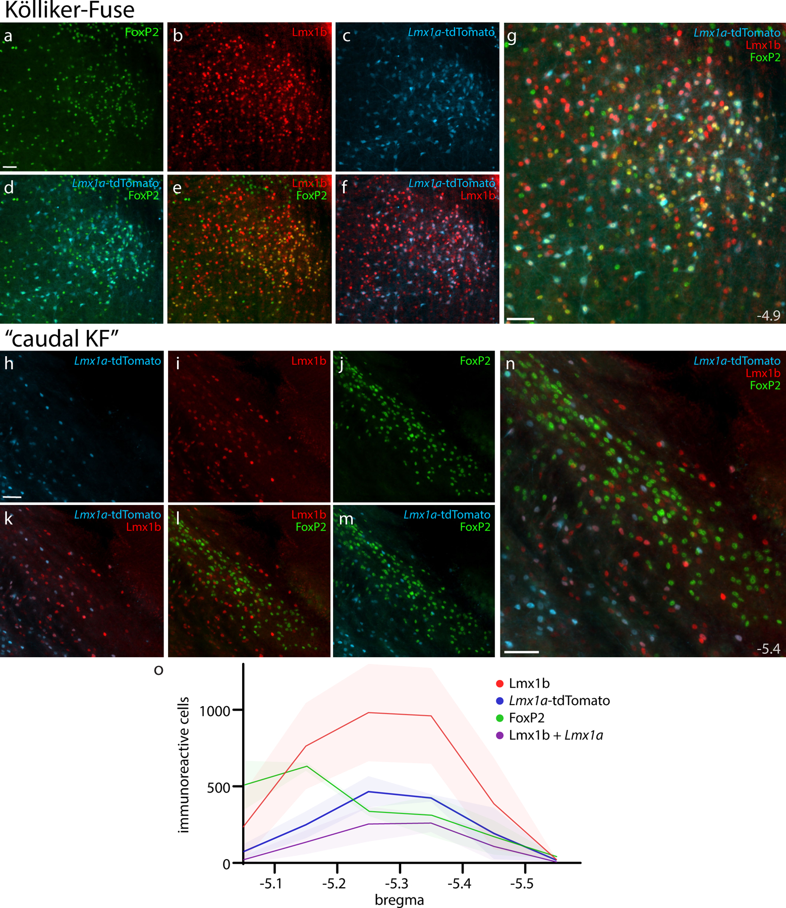
Figure 13. Lmx1a-derived KF neurons and rostral-to-caudal counts across the PB region.

Panels (a–g) show Cre fate-mapping for Lmx1a (tdTomato, pseudocolored ice blue) and immunofluorescence labeling for Lmx1b (red) and FoxP2 (green) in the KF region (ventral to the rostral PB). Panels (h–n) show the lack of Lmx1a Cre-reporter expression in “caudal KF” neurons that contain FoxP2 (ventrolateral to the caudal PB). Approximate level caudal to bregma (in mm) is shown at bottom-right in (g, n). All scale bars are 50 μm. Scale bar in (a) applies to (b–f) and scale bar in (h) applies to (i–m). Bottom graph (o) shows rostral-to-caudal counts of PB neurons expressing the tdTomato Cre-reporter for Lmx1a, FoxP2, or Lmx1b, as well as neurons containing both Lmx1b and Lmx1a Cre-reporter. Counts were averaged at each level (n=3 mice), with variance represented by a standard deviation envelope. Approximate bregma levels are shown on the x-axis.
