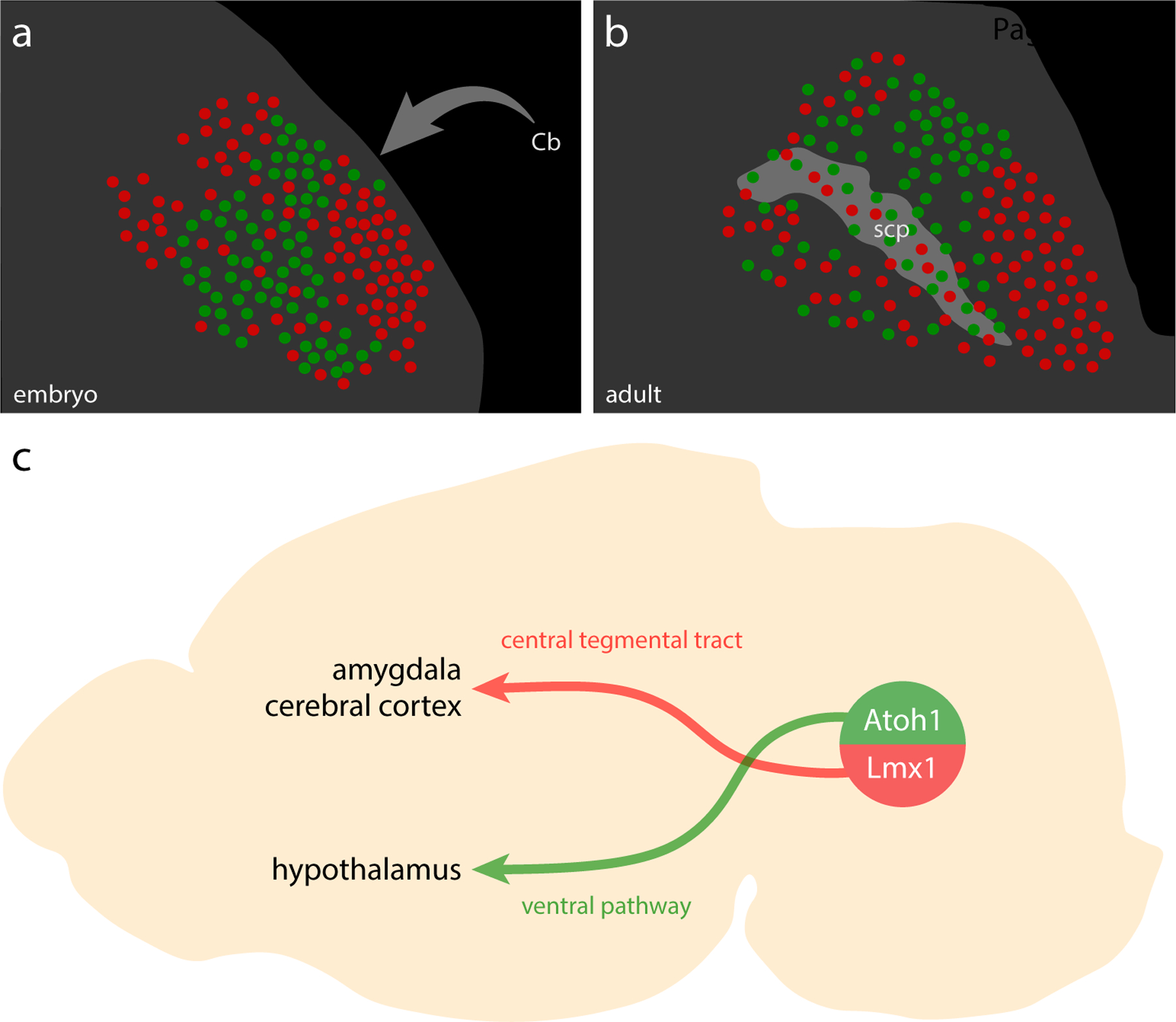
Figure 22. Summary.

(a–b) Illustrated distribution of Atoh1 (green) and Lmx1 (red) PB neurons in late-embryonic (a) and adult (b) mice. Cerebellar axons project through this region and form the scp, dispersing PB neurons. (c) PB neurons in separate Atoh1 and Lmx1 macropopulations project axons through separate pathways to separate forebrain targets. Note that neurons in each macropopulation project to additional brain regions (see D. Huang et al., 2021). Hindbrain projections from ventral populations are not shown.
