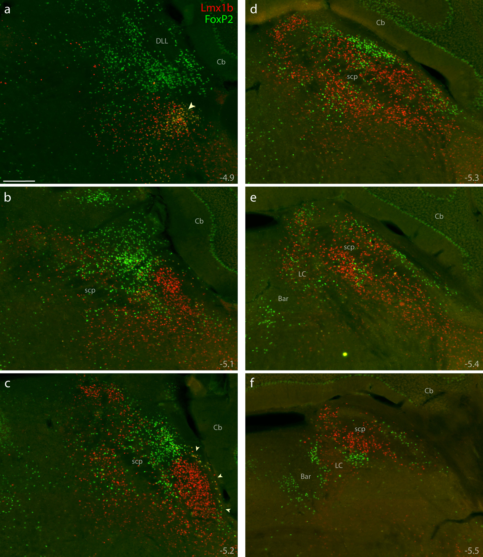
Figure 4. Immunofluorescence labeling for Lmx1b and FoxP2 protein.

Combined immunolabeling for the nuclear transcription factors Lmx1b (red) and FoxP2 (green) identified largely separate distributions across successive, rostral-to-caudal sections through the PB region (a–f). Approximate level caudal to bregma is shown at the bottom-right of each panel (in mm). Lmx1b immunofluorescence labeling was mutually exclusive with FoxP2, except in a rostral cluster of neurons ventral to the PB (arrowhead in a) and in sparse, double-labeled neurons extending back to mid-rostral levels of the PB along the lateral edge of the brainstem (arrowheads in c). Scale bar in (a) is 200 μm and applies to panels (b-f). Other abbreviations: Bar, Barrington’s nucleus; DLL, dorsal nucleus of the lateral lemniscus.
