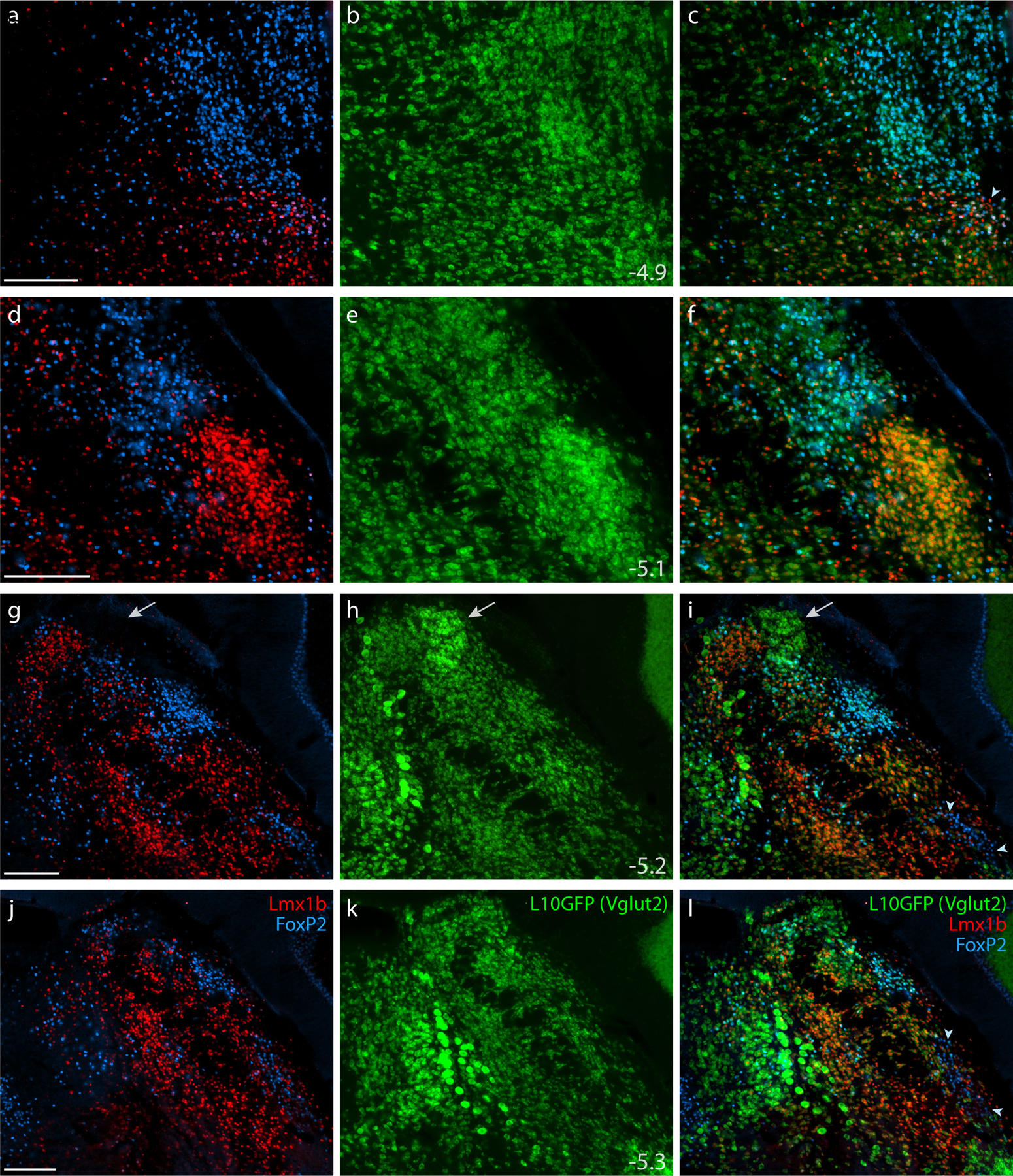
Figure 5. Lmx1b and FoxP2 in a glutamatergic Cre-reporter mouse.

Immunofluorescence labeling identified Lmx1b (red) and FoxP2 (blue) in the PB of mice expressing an L10GFP Cre-reporter (green) for the glutamatergic marker gene Slc17a6 (vesicular glutamate transporter 2, Vglut2). Arrowhead in (c) indicates a cluster of triple-labeled neurons (Lmx1b+FoxP2+L10GFP) located rostrally, in the KF region. White arrows in (g–i) highlight a dorsal cluster of L10GFP-expressing neurons lacking both Lmx1b and FoxP2. Arrowheads in (i) and (l) indicate a ventrolateral cluster of neurons labeled for FoxP2 in the “caudal KF.” Approximate bregma levels are shown at bottom-right in the center column (in mm). All scale bars are 200 μm and apply to other panels in their respective rows.
