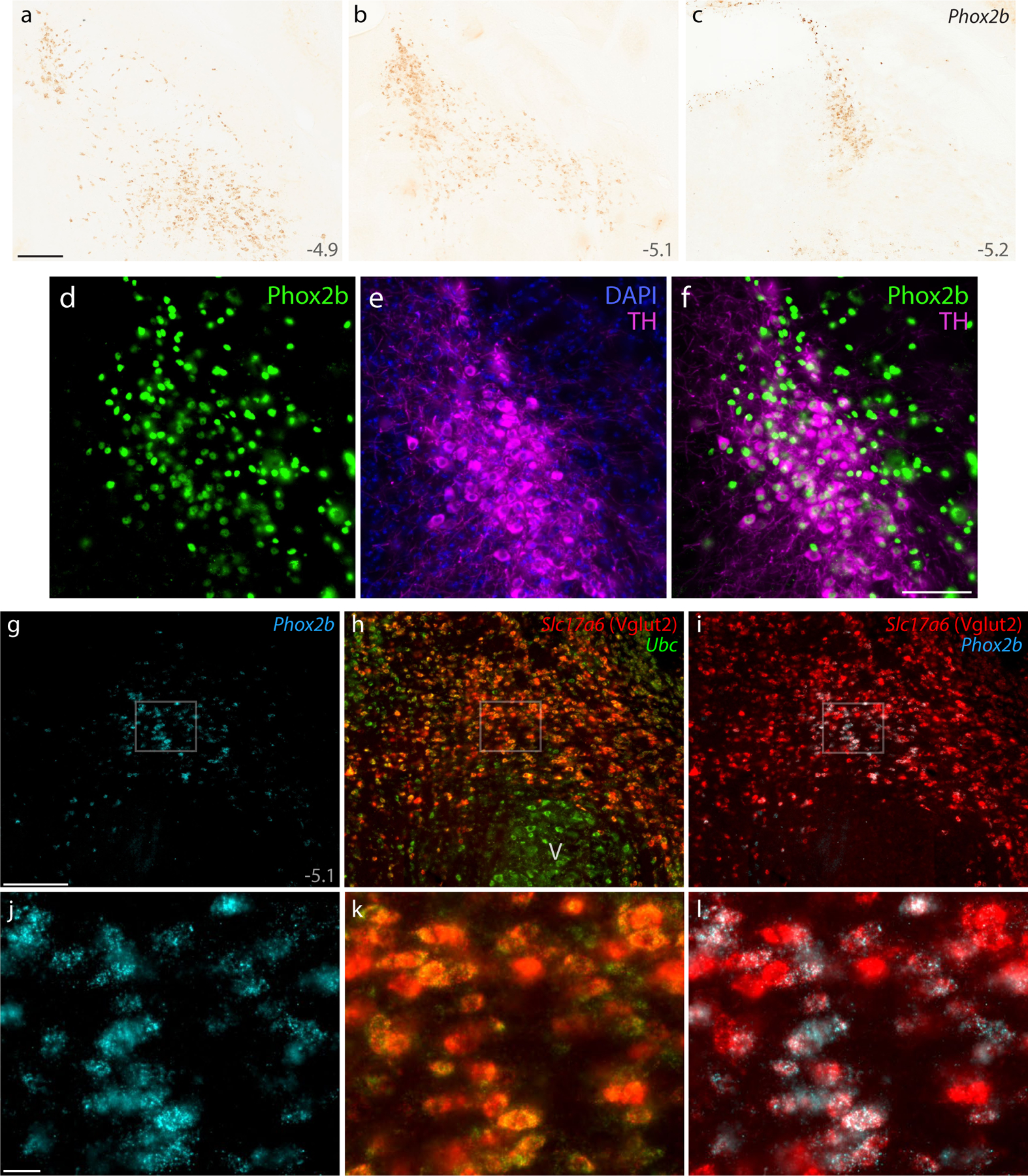
Figure 7. Phox2b identifies glutamatergic and catecholaminergic populations ventral and medial to the PB.

DAB in situ hybridization revealed Phox2b mRNA, shown at three rostral-to-caudal levels (a–c). Approximate level caudal to bregma is shown at the bottom-right of each panel (in mm). (d–f) Immunofluorescence labeling for Phox2b (green) and tyrosine hydroxylase (TH, magenta) to identify LC neurons. (g–i) FISH identified co-localization between Phox2b mRNA (ice-blue) and the glutamatergic marker Slc17a6 mRNA (h, red) ventral to a mid-level of the PB (bregma −5.1 mm). Ubc mRNA (green) is shown for neuroanatomical background. (j–l) Blow-ups of the highlighted region in (g–i) show the ubiquitous co-localization of Slc17a6 mRNA in Phox2b-expressing neurons in the supratrigeminal region. All scale bars are 200 μm and apply to related panels. Other abbreviations: V, motor trigeminal nucleus.
