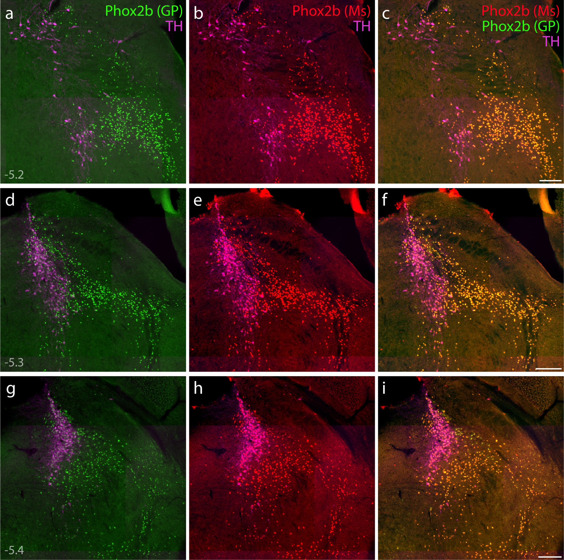
Figure 8. Immunofluorescence labeling for Phox2b.

(a–i) Direct comparison of immunofluorescence labeling between guinea pig polyclonal [green, “Phox2b (GP)”; (Nagashimada et al., 2012)] and mouse monoclonal [red, “Phox2b (Ms)”; sc-376997] anti-Phox2b antisera, combined with TH immunofluorescence (magenta) to identify LC neurons. In addition to confirming antibody specificity, labeling Phox2b across three rostral-to-caudal levels of the PB region highlighted the extensive population of non-LC (glutamatergic) Phox2b neurons, which form an observer-independent ventromedial border for the PB. Approximate bregma levels are shown at bottom-left in (a, d, g). Scale bars in (c, f, i) are 200 μm, and each scale bar applies to other panels in the same row.
