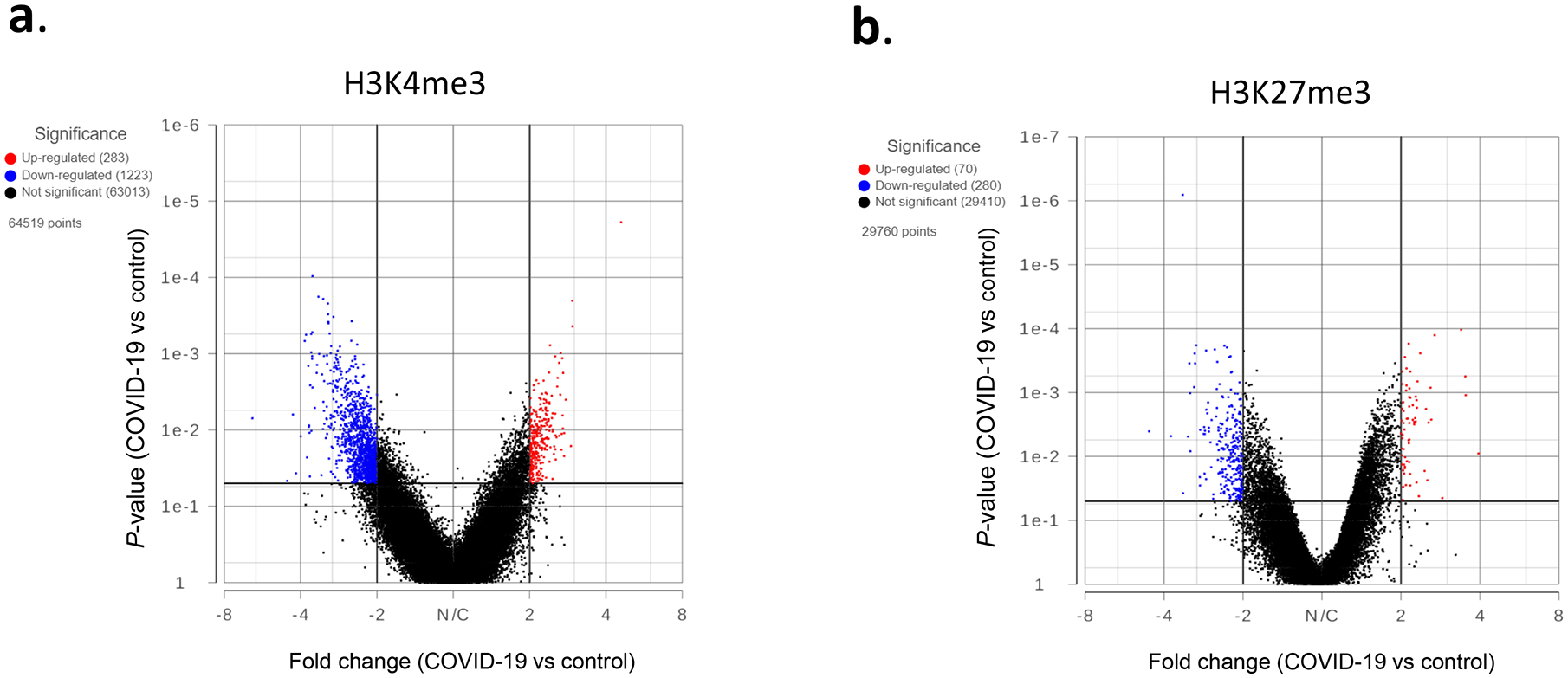
Figure 2. Genes with altered histone marks.

H3K4me3 (a) and H3K27me3 (b) signals within 5kb upstream and downstream of TSS in PBMCs of 5 controls and 5 COVID-19 patients were quantified by Partek software using normalized sequencing counts. Red dots represent the genes with significantly increased histone mark in the COVID-19 samples, while blue dots are genes with reduced mark.
