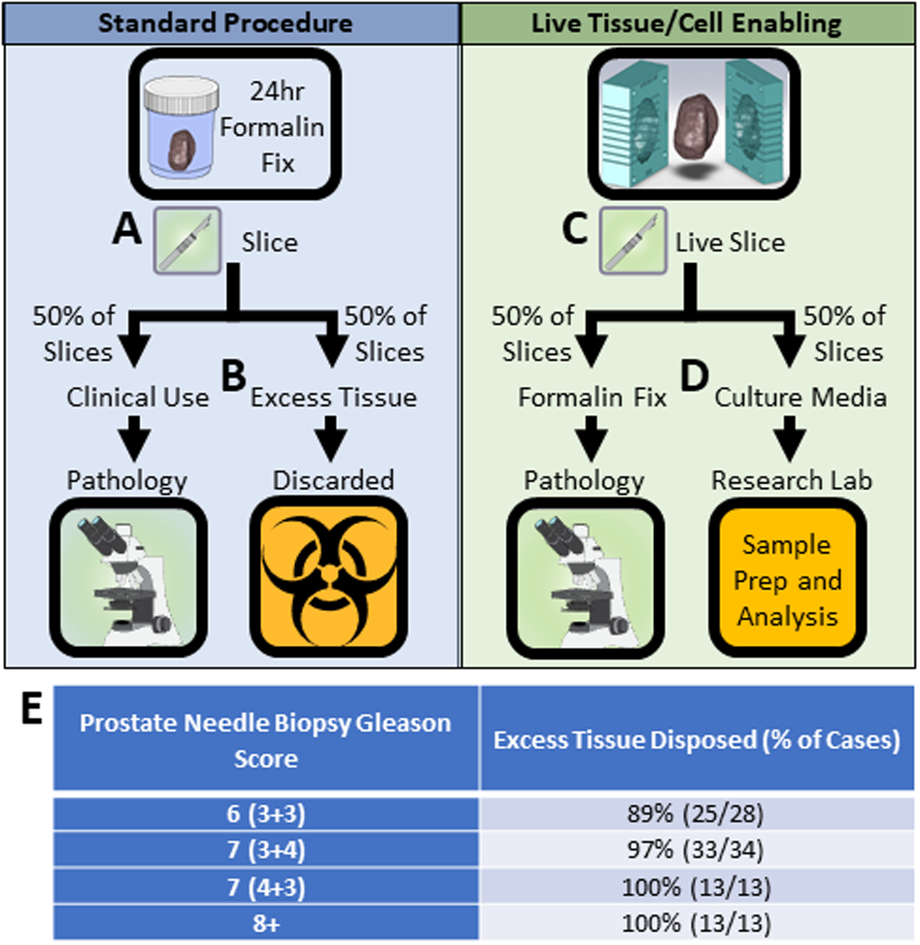
Figure 1: Live tissue slicing enables standard of care histopathology as well as live cell functional and molecular readouts in a wet lab.

(A) In standard hospital procedure, prostate tissue is fixed in formalin for 24 hours then cut into 4mm slices. (B) Tissue needed for standard of care histology (~50% of tissue slices) is sent to hospital pathology, excess tissue is discarded. (C) In live tissue/cell enabled protocols, prostate tissue is placed in a slicing mold and cut into 4mm slices before formalin fixation. (D) Tissue needed for standard of care histology (~50% of tissue slices) is formalin fixed and sent to hospital pathology, excess tissue is placed in live culture media and remains viable for sample processing and analysis at the research wet lab. (see Figure 2). (E) Prostatectomy cases (n=88) at University of Wisconsin – Hospital and Clinics in which excess prostate tissue was disposed of after clinical analyses were complete.
