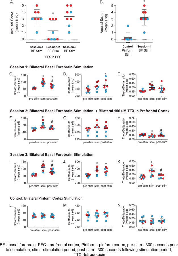
Figure 5.
Inactivation of prefrontal cortex attenuates electroencephalographic, physiologic, and behavioral arousal induced by bilateral electrical stimulation of basal forebrain during sevoflurane anesthesia. Each red (female) and blue (male) dots show the individual rat data. A, Comparison of behavioral arousal score after bilateral electrical stimulation of basal forebrain with or without concurrent inactivation of prefrontal cortex. As compared to the bilateral electrical stimulation of basal forebrain without prefrontal inactivation (session 1), bilateral electrical stimulation of basal forebrain with concurrent prefrontal inactivation (session 2) caused a significant decrease in the arousal score. Repeat of session 1 (i.e., session 3) showed the arousal score to be significantly higher than that observed in session 2 and reached the levels observed during session 1. B, Behavioral arousal score after bilateral electrical stimulation of basal forebrain (session 1) were significantly higher than the arousal score after bilateral electrical stimulation of piriform cortex (anatomical control). Bilateral electrical stimulation of piriform cortex did not produce behavioral arousal as in session 1 rats. C-E, Session 1 - bilateral electrical stimulation of basal forebrain in sevoflurane-anesthetized rats increased respiration rate (C), heart rate (D) and theta/delta ratio (E). The pre-stimulation data were quantified 300 s prior to stimulation (pre-stim) and the post-stimulation data were quantified 300 s after the stimulation (post-stim). F-H, Session 2 - bilateral basal forebrain stimulation in the presence of tetrodotoxin (TTX) in prefrontal cortex produced a significant increase in respiration rate (F), but no significant change was observed in heart rate (G). Theta/delta ratio showed a significant decrease during electrical stimulation (H). I-K, Session 3 - bilateral electrical stimulation of basal forebrain in the same rats used in sessions 1 and 2 produced increase in respiration rate (I), heart rate (J), and theta/delta ratio (K). A video showing representative behavior after stimulation of 1) basal forebrain, and 2) after stimulation of basal forebrain along with concurrent inactivation of prefrontal cortex is provided in the Supplemental Digital Content, Video 2. L-N, Bilateral electrical stimulation of piriform cortex during sevoflurane anesthesia did not produce any statistical change in respiration rate (L), heart rate (M), or theta/delta ratio (N). A linear mixed model controlling for sex was used for within-rat statistical comparisons while a linear regression controlling for sex was used for between-rat (B) statistical comparisons. Post-hoc comparisons were Tukey corrected. The P values are shown at < .05 but the actual P values are reported in the main text and in the Supplemental Digital Content, Tables S4–S8. For panel A: *Compared to session 1. For panel B: *Compared to Control. For panels C-N: *Compared to pre-stimulation, #compared to stimulation.

