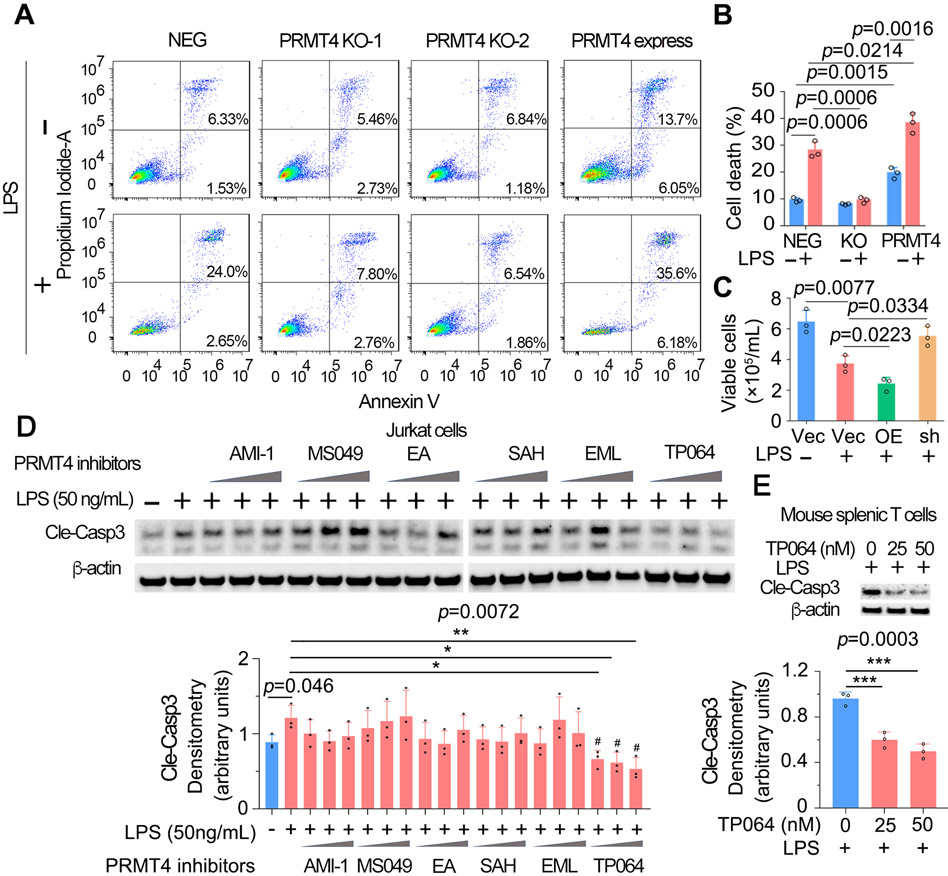
Fig. 5. High protein level of PRMT4 causes lymphocyte death.
A, B. FACS analysis of apoptosis in PRMT4 KO or overexpressed Jurkat cells with or without LPS treatment. Data of A were quantitated in B. C. Lenti-PRMT4 or shRNA particles were delivered intratracheally into the mouse. Mouse splenic T cells were isolated and treated with LPS for 18 h, viable cells were counted. “Vec” denotes vector; “OE” denotes PRMT4 overexpression; “sh” denotes PRMT4 shRNA. D. Jurkat cells were treated with LPS and a range of PRMT4 inhibitors as indicated for 3 h. Cell lysates were analyzed for cleaved caspase 3. Relative expression of cleaved caspase 3 in each group were plotted in the lower panel. Independent experiments n=3. E. Isolated mouse splenic T cells were treated with LPS and TP064, cleaved caspase 3 was immunoblotting analyzed and plotted in the lower panel. Independent experiments n=3. “*” denotes p=0.05-0.01, “**” denotes p=0.01-0.001, “***” denotes p=0.001-0.0001.

