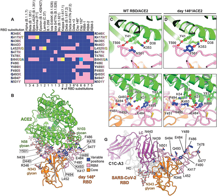
Fig. 1. Structure of intra-host evolved RBD bound to human ACE2.
(A) Key RBD substitutions discussed in the manuscript and the SARS-CoV-2 variants that contain them. (B) Day 146* RBD/ACE2 ectodomain X-ray crystal structure. RBD residues that are mutated in variants discussed in the text are shown. Boxed residues are mutated in the day 146* RBD as compared to the Wuhan-Hu-1 (wild-type) SARS-CoV-2 RBD. RBM: receptor-binding motif. *In addition to the mutations that are shown, the Delta +3 variant contains an additional RBD mutation that is not shown in the schematic diagram (see table S2). (C) Wild-type RBD ACE2 contacts near N501RBD (PDB ID: 6M0J) (2). (D) Day 146* RBD contacts near Y501RBD. (E) Wild-type SARS-CoV-2 RBD ACE2 interactions near Q493RBD. (F) Day 146* RBD interactions near K493RBD. (G) cryo-EM structure of the SARS-CoV-2 RBD bound to the C1C-A3 antibody Fab. RBD residues discussed in the text are labeled.

