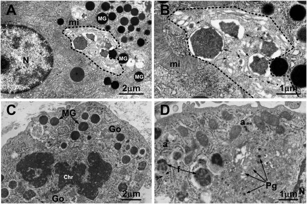
Fig. 1.
(A,B) Transmission electron micrographs of a pancreatic acinar cell from a mouse killed 8 h after pilocarpine administration to induce secretion of granules. Many mature granules (MG in A) are present in the cytoplasm outside of the progranule zone (indicated by dashed line), which is limited by the outermost of the Golgi cisternae, the mature granules, and the rough endoplasmic reticulum. Within the progranule zone are several membrane-delimited structures of various sizes (arrows in B), containing electron dense content that is more variegated in appearance than that in the mature granules; the smallest of these structures are interpreted as progranules and the larger ones may represent the products of fusion of progranules. mi mitochondrion; N nucleus. Bars: (A) 2 μm; (B) 1 μm. (A and B are reproduced in modified form from S. Lew, I. Hammel and S. J. Galli. Cell Tissue Res. 278: 327-336, 1994, with the permission of the publisher.) (C,D) Transmission electron micrographs of mast cells in the subcutaneous tissue of a 35 mm rat embryo (C) or a newborn rat (D). In (C), a mast cell in mitosis exhibits some mature secretory granules (MG) with homogeneously electron dense content. Chr chromatin. Numerous progranules (Pg in D) are present in the Golgi zone (Go in C). In (D), several progranules appear to have aggregated at “a” and larger structures (f), representing immature granules, appear to contain progranules in various stages of fusion; the arrows near f indicate the close association of the rough endoplasmic reticulum with one of the immature granules. N nucleus. Bars: (C) 2 μm; (D) 1 μm. (C & D are figures 3 & 4, reproduced in modified form from J. W. Combs. J Cell Biol. 31: 563-75, 1966, with the permission of the publisher.)

