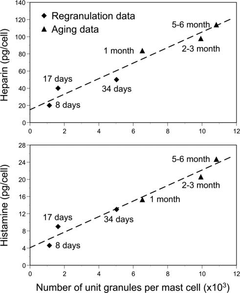
Fig. 4.
Heparin (upper panel) and histamine (lower panel) content of rat peritoneal mast cells as a function of number of unit granules per mast cell. “Regranulation data” (indicated by solid diamonds) were derived from rats sacrificed for analysis of peritoneal mast cells by transmission electron microscopy 8, 17 or 34 days after the mast cells were induced to degranulate in vivo by 4 injections (i.p.) of polymyxin B (mg/kg body weight of 2.8 on day 1, 5.7 and days 2 and 3 and 12.0 and day 4). “Aging data” (indicated by solid triangles) were derived from control rats (not injected with mast cell activating agents) sacrificed for analysis of peritoneal mast cells by transmission electron microscopy at 1, 2-3 or 5-6 months of age. (Data in upper panel are from P. G. Krüger and D. Lagunoff. Int Arch Allergy Appl Immunol. 65: 291-299, 1981a and P. G. Krüger and D. Lagunoff. Int Arch Allergy Appl Immunol. 65: 278-290, 1981b, with the permission of the publisher: S. Karger, Basel; figure in lower panel is reproduced from I. Hammel, D. Lagunoff, and P. G. Krüger. Exp Cell Res. 184: 518-523, 1989, with the permission of the publisher.)

