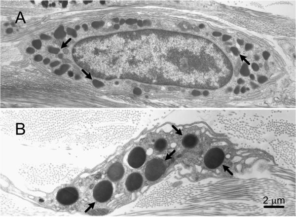
Fig. 5.

Dermal mast cells from (A) a C57BL/6 wild type mouse and (B) a C57BL/6-Lystbg/Lystbg (“beige”) mouse. The secretory granules (some indicated by arrows) in the beige mouse mast cell are much larger and fewer in number than those in the wild type mouse mast cell. Bar (for A and B): 2 μm.
