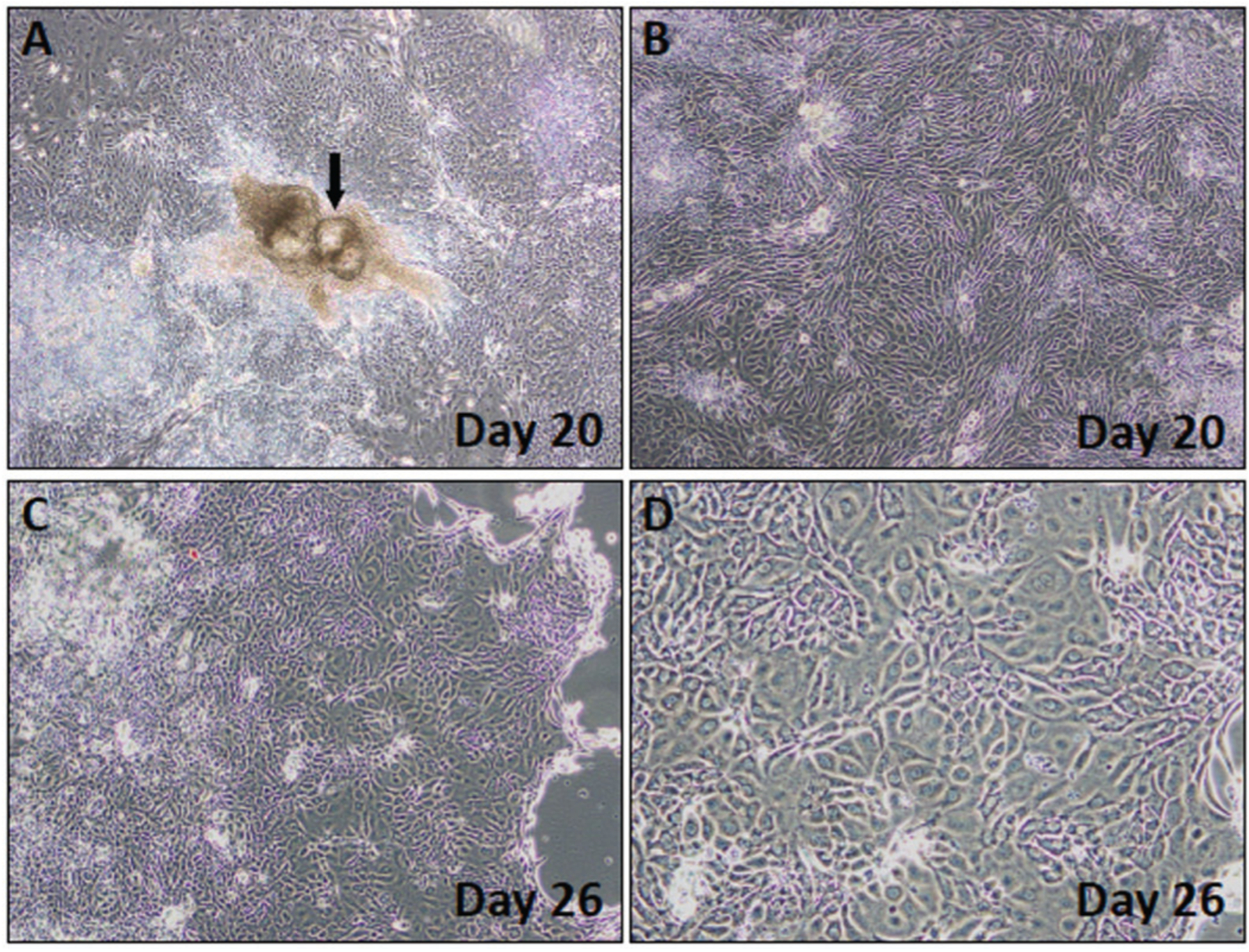
Figure 4.

Phase-contrast images showing colony morphologies observed during the selection phase. (A,B) Day 20 colonies. Arrow in (A) points to a colony with abnormal morphology. (B) Day 20 colony with the expected cobblestone cell morphology (C,D) Day 26 cells with emerging keratinocyte morphology. Note the shiny edge in the colony shown in (C), a sign that the cells are ready to be passaged.
