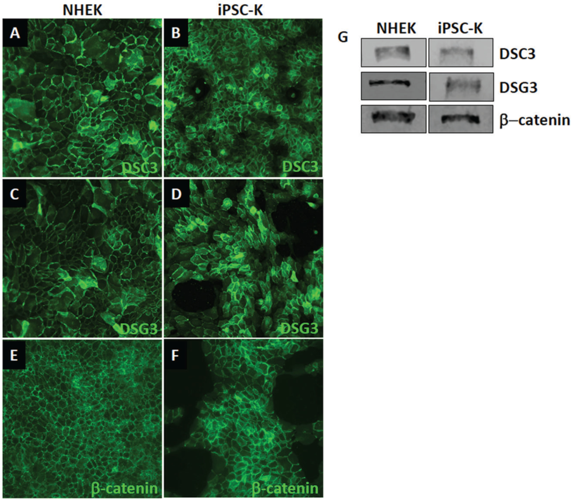
Figure 6.

Expression of keratinocyte markers in iPSC-K. iPSC-K and primary human epidermal keratinocytes were exposed to 1.3 mM Ca2+ for 48 hr. (A-F) Immunofluorescence staining and (G) Western blot analysis demonstrates normal expression and localization of desmosomal proteins (DSC3, DSG3) and an adherens junction protein (β-catenin). Antibodies used for immunofluorescence staining are: DSG3 (clone 5H10; courtesy of Dr. James K Wahl III, PhD, University of Nebraska Medical Center, Lincoln, NE), DSC3 (Progen cat. no. 61093), and β-catenin (Santa Cruz cat. no. sc-7963). Antibodies used forlotting are: DSG3 (Invitrogen cat. no. MA5–16025), DSC3 (Progen cat. no. 61093), and β-catenin (Santa Cruz cat. no. sc-7963).
