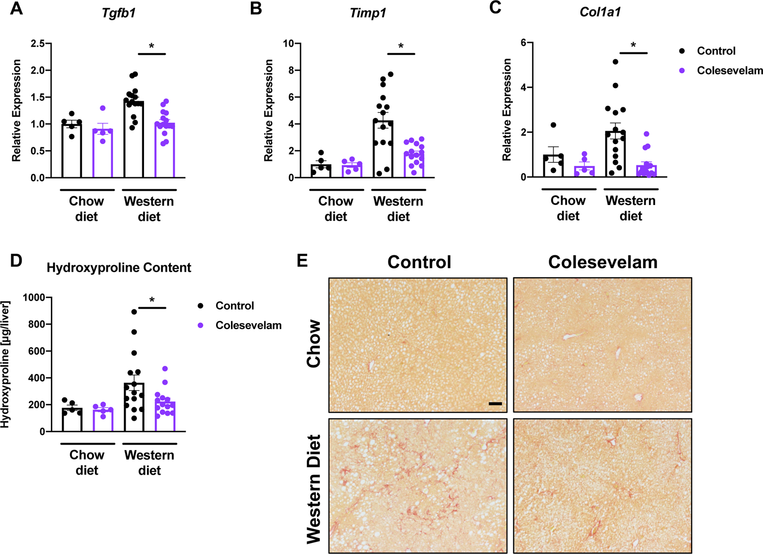
Figure 3. Colesevelam decreases Western diet-induced hepatic fibrosis in microbiome-humanized mice.

Microbiome-humanized C57BL/6 mice were fed a chow diet (n=5) or Western diet (n=14–15) with or without colesevelam for 20 weeks. (A-C) Hepatic mRNA expression of (A) Tgfb1, (B) Timp1, and (C) Col1a1. (D) Hepatic hydroxyproline content. (E) Representative liver sections after Sirius Red staining (bar size = 100 μm). Results expressed as mean ± s.e.m. P values are determined by Mann-Whitney test (A, C) or One-way ANOVA with Holm’s post-hoc test (B, D). *P<0.05. Tgfb1, transforming growth factor-beta 1; Timp1, tissue inhibitor of metalloproteinase-1.
