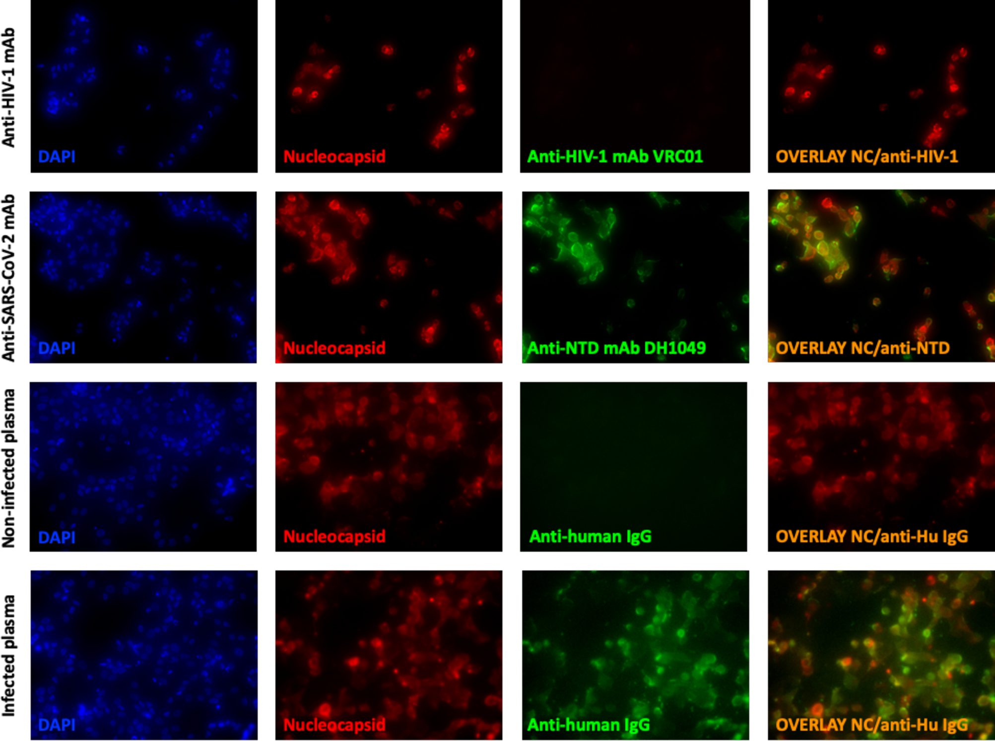
Figure 2. Fluorescent microscopy showing antibody binding SARS-CoV-2-infected cells.

Infected Vero E6 cells were stained intracellularly with an anti-SARS-CoV-2 Nucleocapsid antibody (Red), on the surface with controls (anti-HIV-1 monoclonal antibody, VRC01, or plasma from an uninfected individual) or SARS-CoV-2 specific antibody (anti-SARS-CoV-2 NTD mAb, DH1049, or sera from a SARS-CoV-2-infected individual) (green). Nucleated cells were identified using DAPI (blue) and an overlay of anti-SARS-CoV-2 Nucleocapsid and anti-SARS-CoV-2 antibodies bound to the surface of cells is shown (right column). Cells were visualized and pictures taken at 100x.
