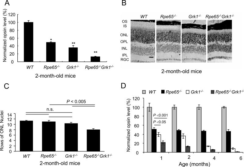
Figure 2.
Effect of deletion of Rpe65 and Grk1on retinal opsin levels and morphology. A, Opsin levels were calculated from rhodopsin that formed upon the addition of 11-cis retinal. Data were generated from 2-month-old cyclic-light reared mice. The relative opsin levels in each strain were normalized to the average of wild type opsin concentration and shown as a fraction of the wild type. B, Retinal morphology of 2-month-old WT, Rpe65−/−, Grk1−/− and Rpe65−/−Grk1−/− mice. Light micrographs of paraffin-embedded retinae sections from the superior central region of the retina. While morphology of the inner retina was unaffected by the different genotypes at that resolution, the outer retina showed shortening of the inner and outer segments progressing in severity from the Rpe65−/− < Grk1−/− << Rpe65−/−Grk1−/− (scale bar: 30 μm). Abbreviations: INL, inner nuclear layer; IPL, inner plexiform layer; IS, inner segments; ONL, outer nuclear layer; OPL, outer plexiform layer; OS, outer segments; RGC, retinal ganglion cells. C, Bar graph representing rows of photoreceptor cell nuclei counted in the superior central region of the retinae of 2-month-old mice from the 4 strains; mean ± SEM; n=5. The cell loss was significant for the Rpe65−/−Grk1−/− mice. D, Opsin levels with age, assayed as for panel A. The relative opsin levels in each strain were normalized to the average of wild type opsin concentration at each age, and shown as the fraction of wild type. Grey bar, WT mice; black bar, Rpe65−/− mice; white bar, Grk1−/− mice; hatched bar, Rpe65−/−Grk1−/− mice.

